3D-Viewer CLICK HERE
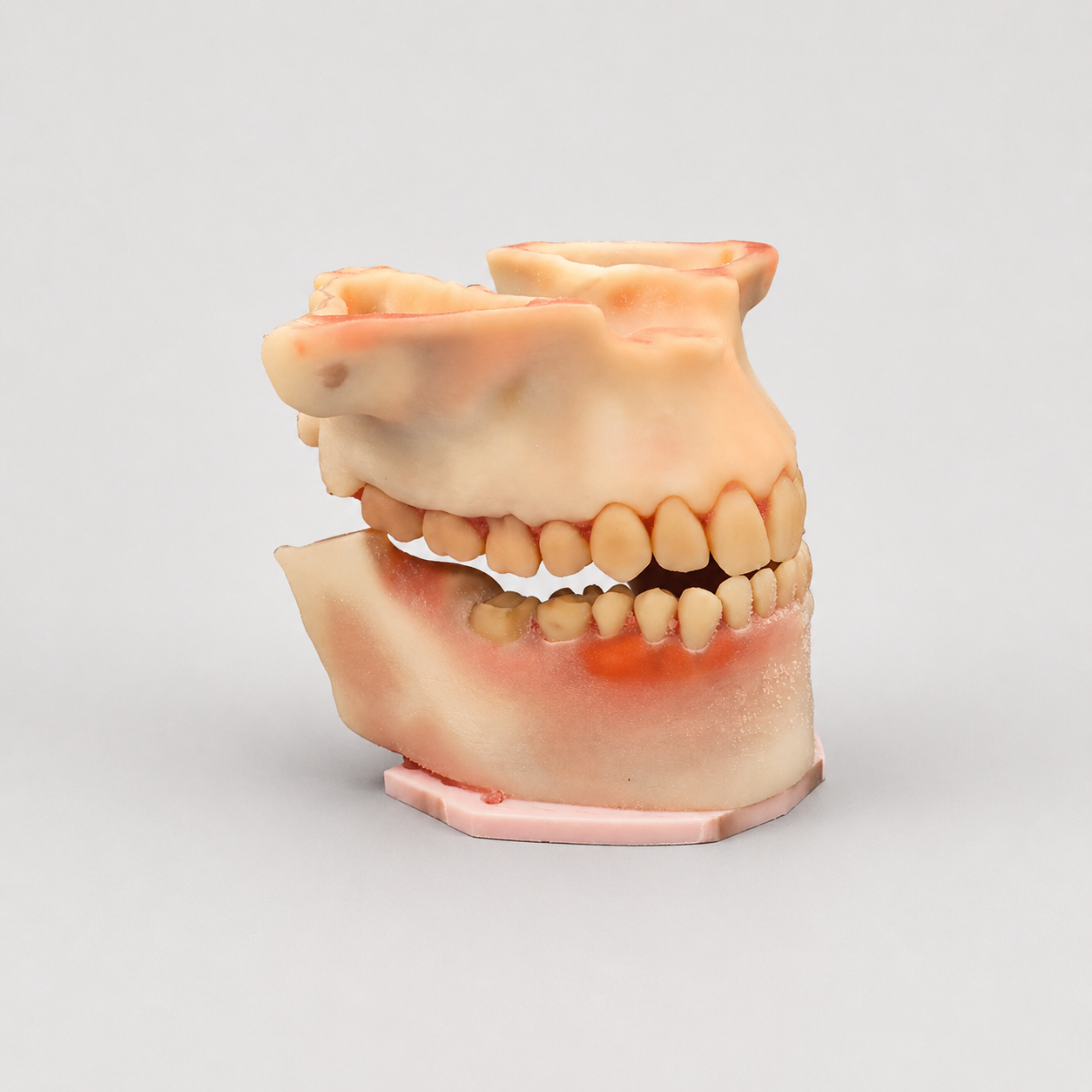
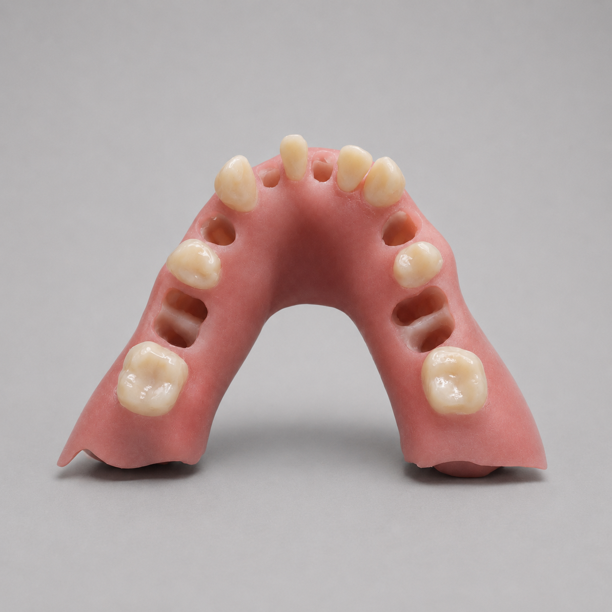
Train advanced maxillary oral surgery procedures with our Maxillary Extraction Model with Third Molars, Greater Palatine, and Incisive Canal Nerve Anatomy designed for dental schools, CE courses, OMS programs, implant educators, and clinicians.
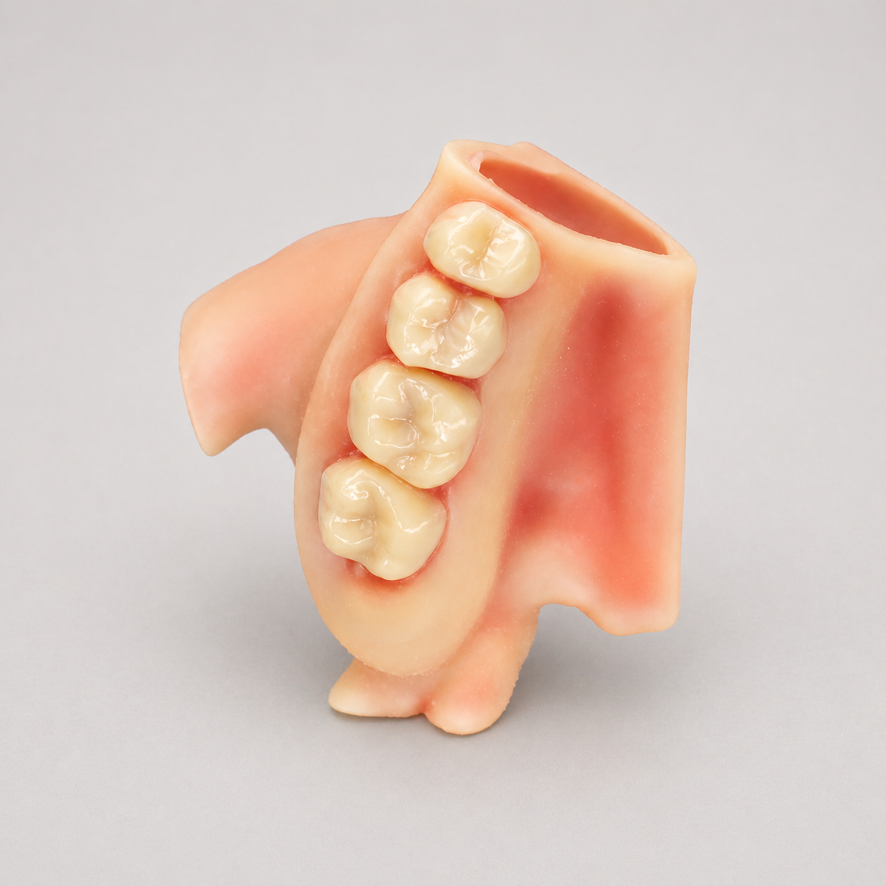
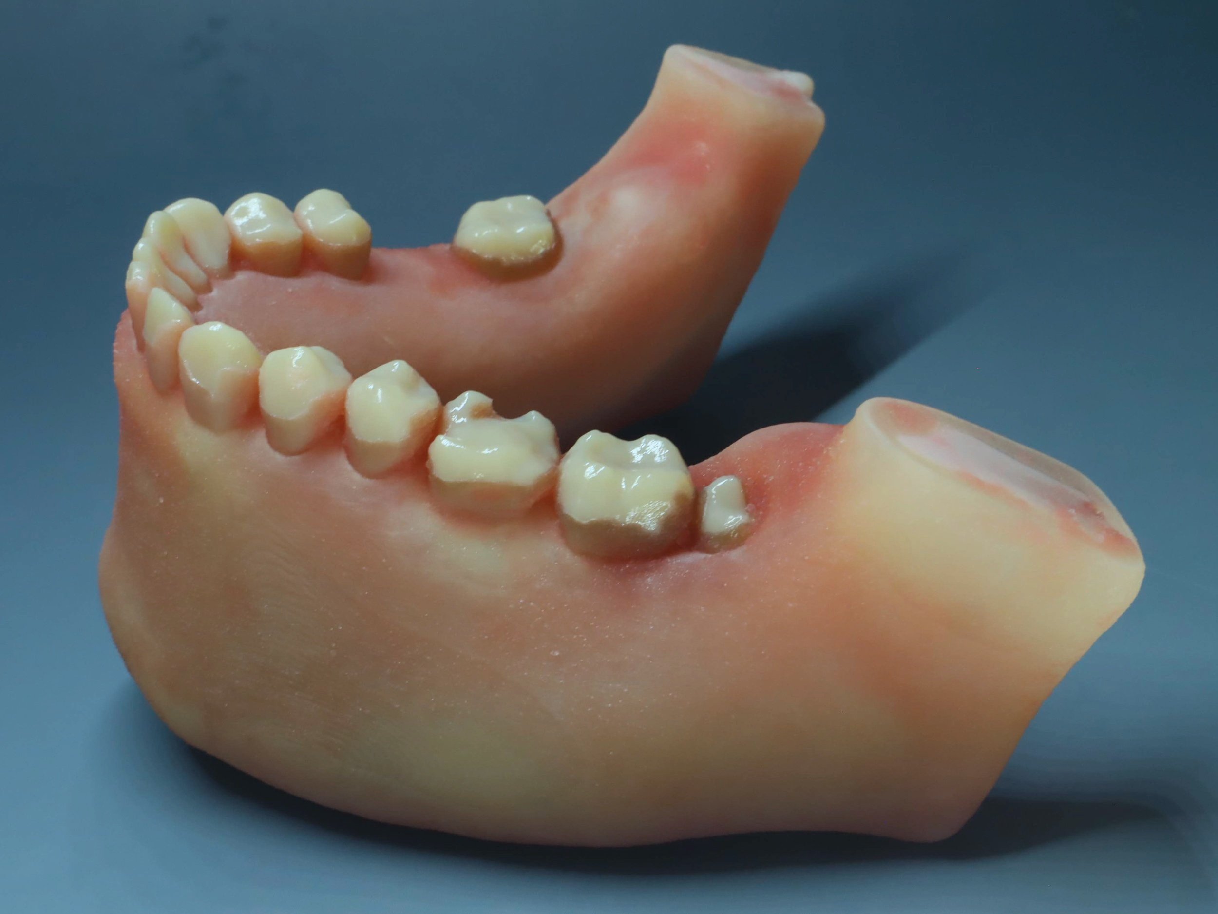
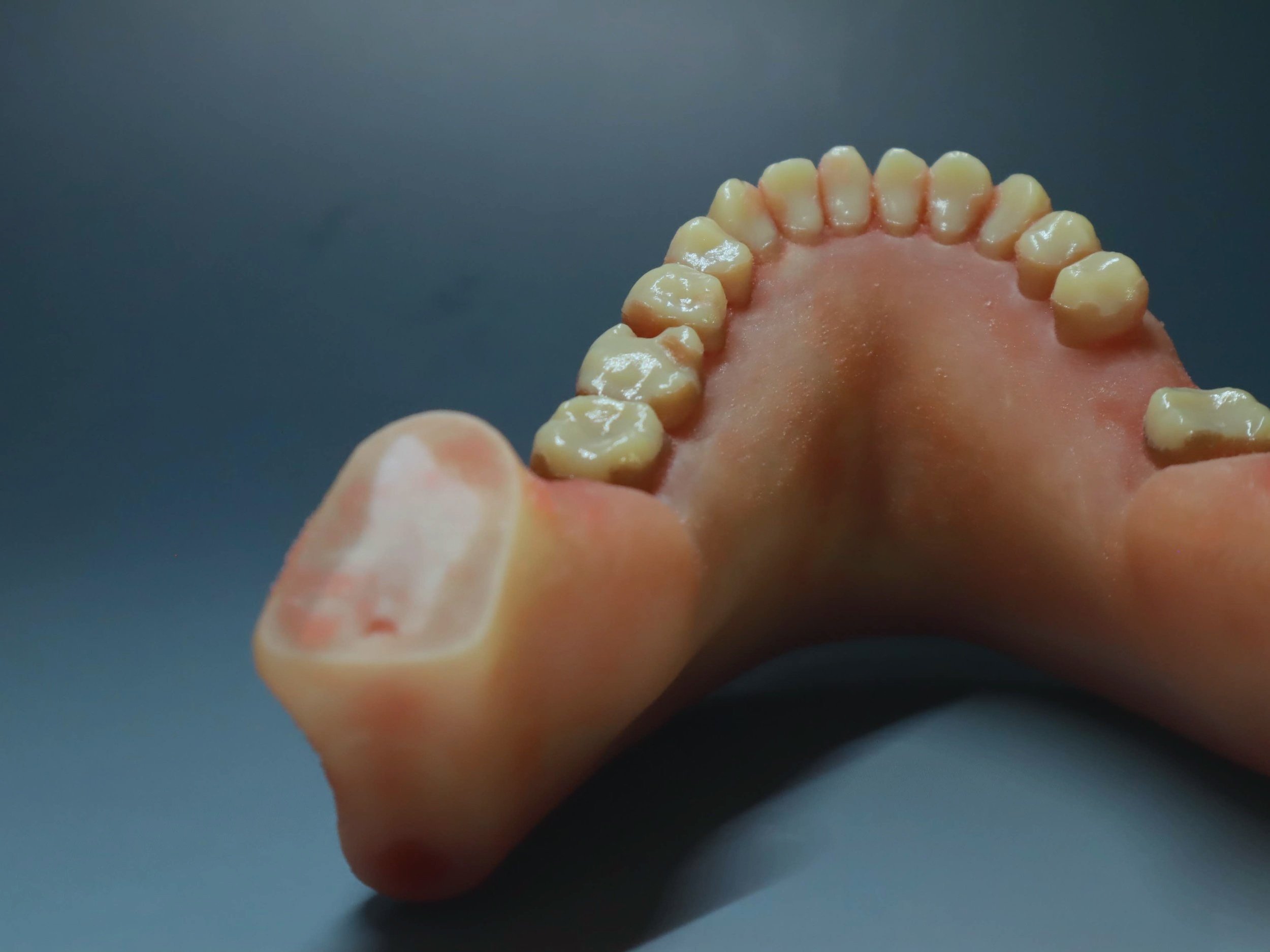
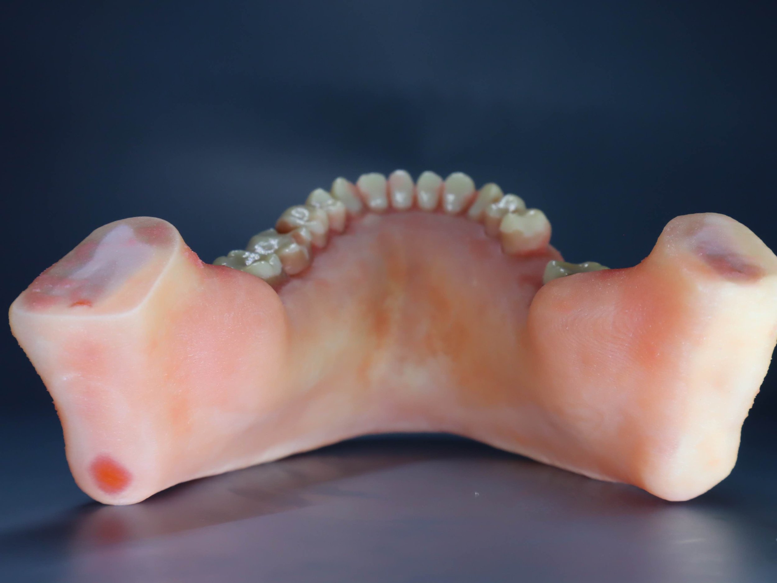
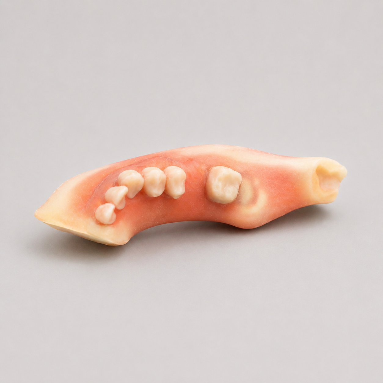
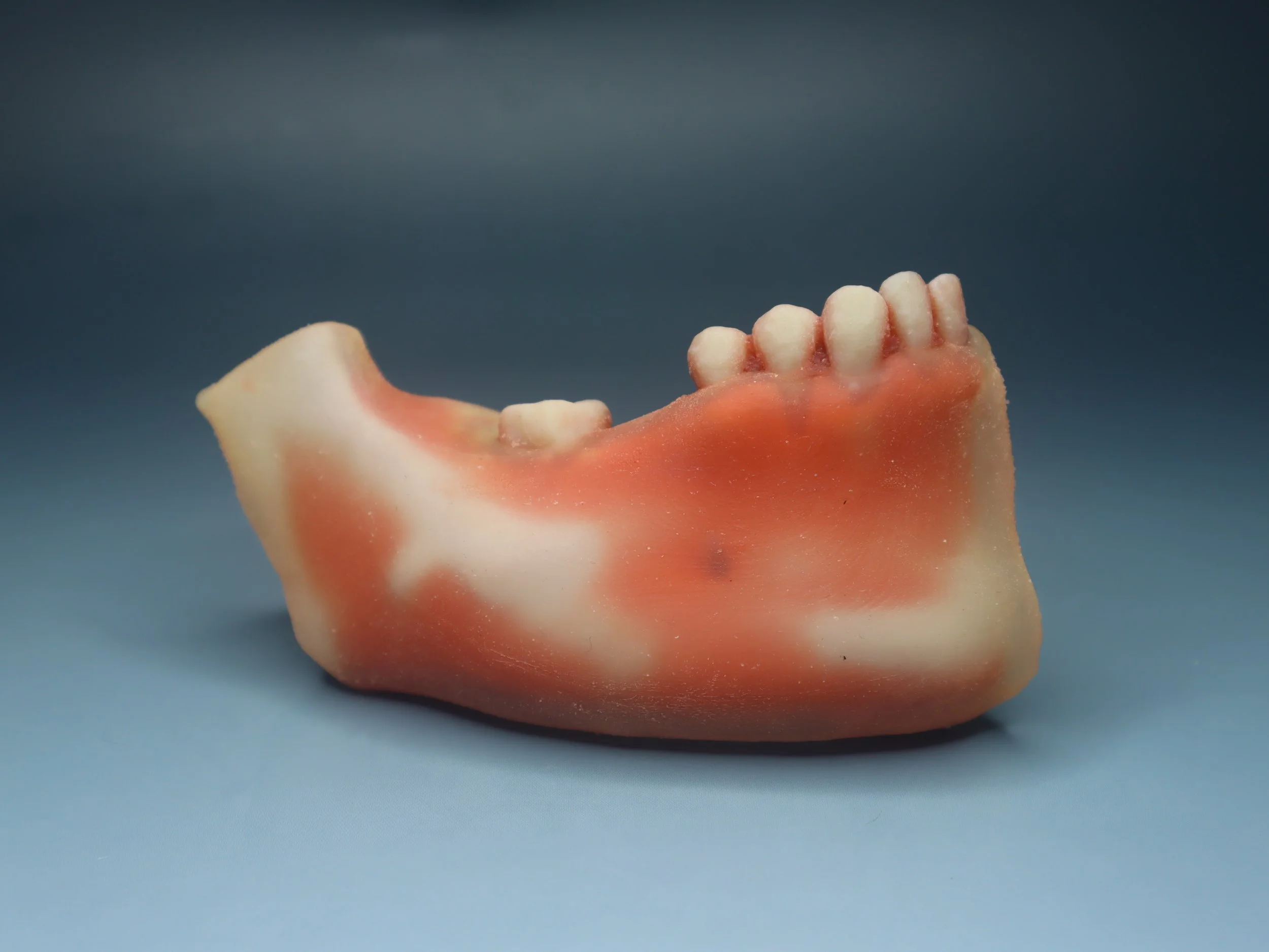
This model simulates realistic maxillary anatomy for extractions, flap design, implant planning, sinus-related procedures, and anatomical landmark training.
Includes third molars, sinus proximity, palatal nerve landmarks, and soft tissue options for comprehensive hands-on education.
Best For
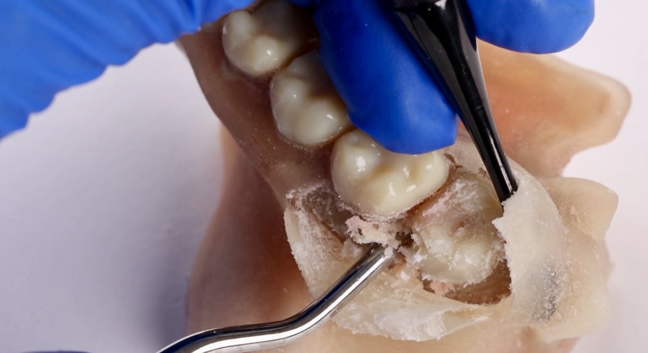
• Maxillary third molar extraction training
• Flap elevation and access design
• Sectioning and osteotomy practice
• Implant placement planning
• Sinus proximity case training
• Suturing techniques
• Palatal anatomy education
Key Features
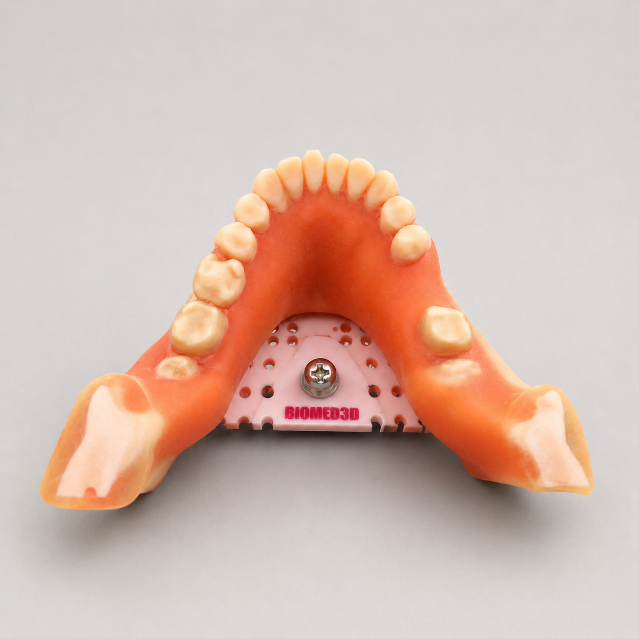
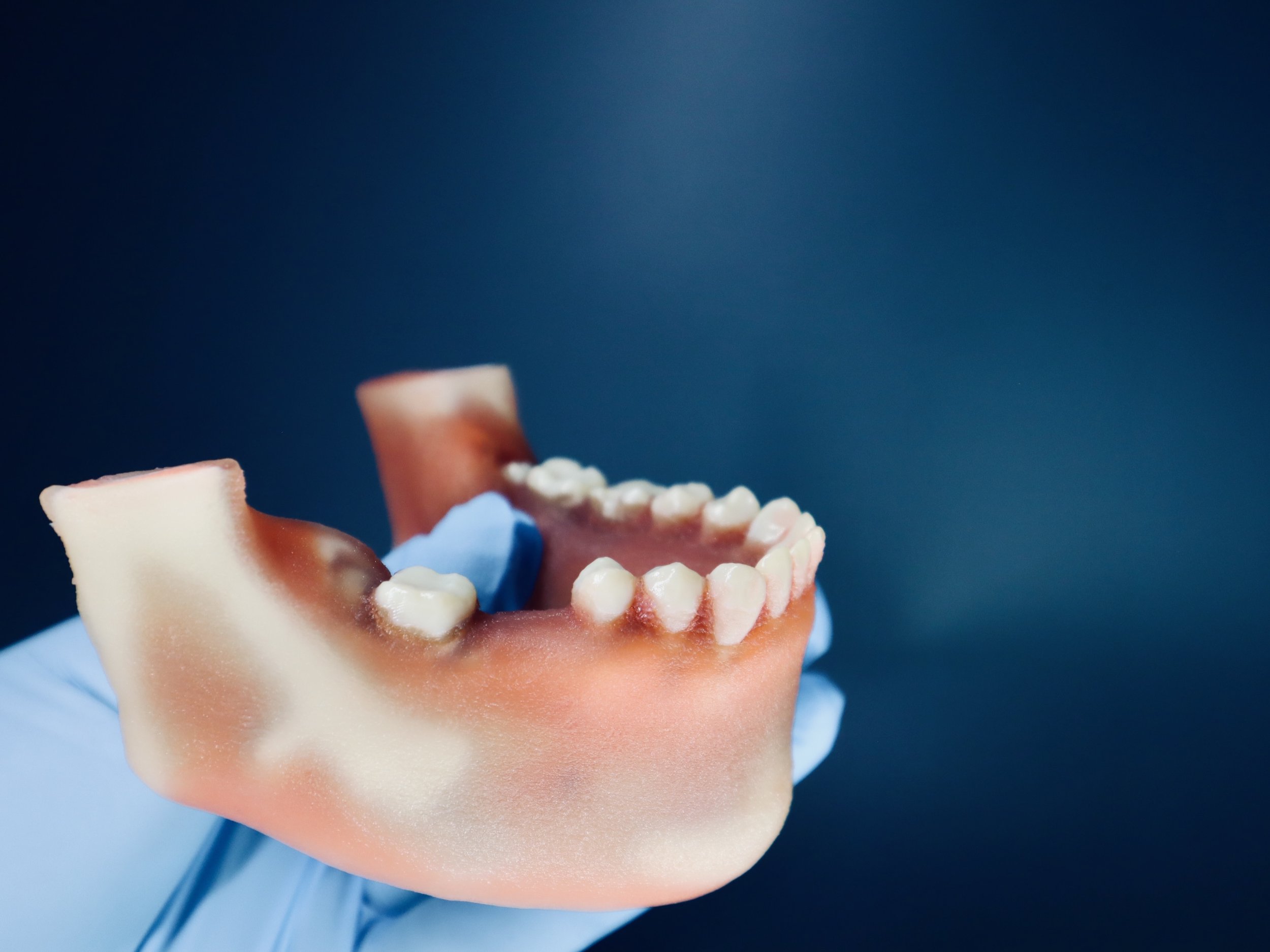
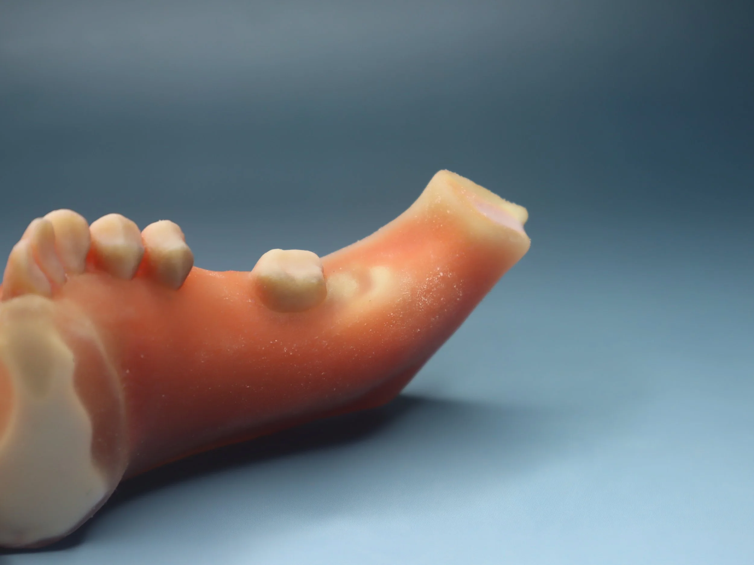
• Realistic maxillary jaw anatomy
• Includes upper third molars
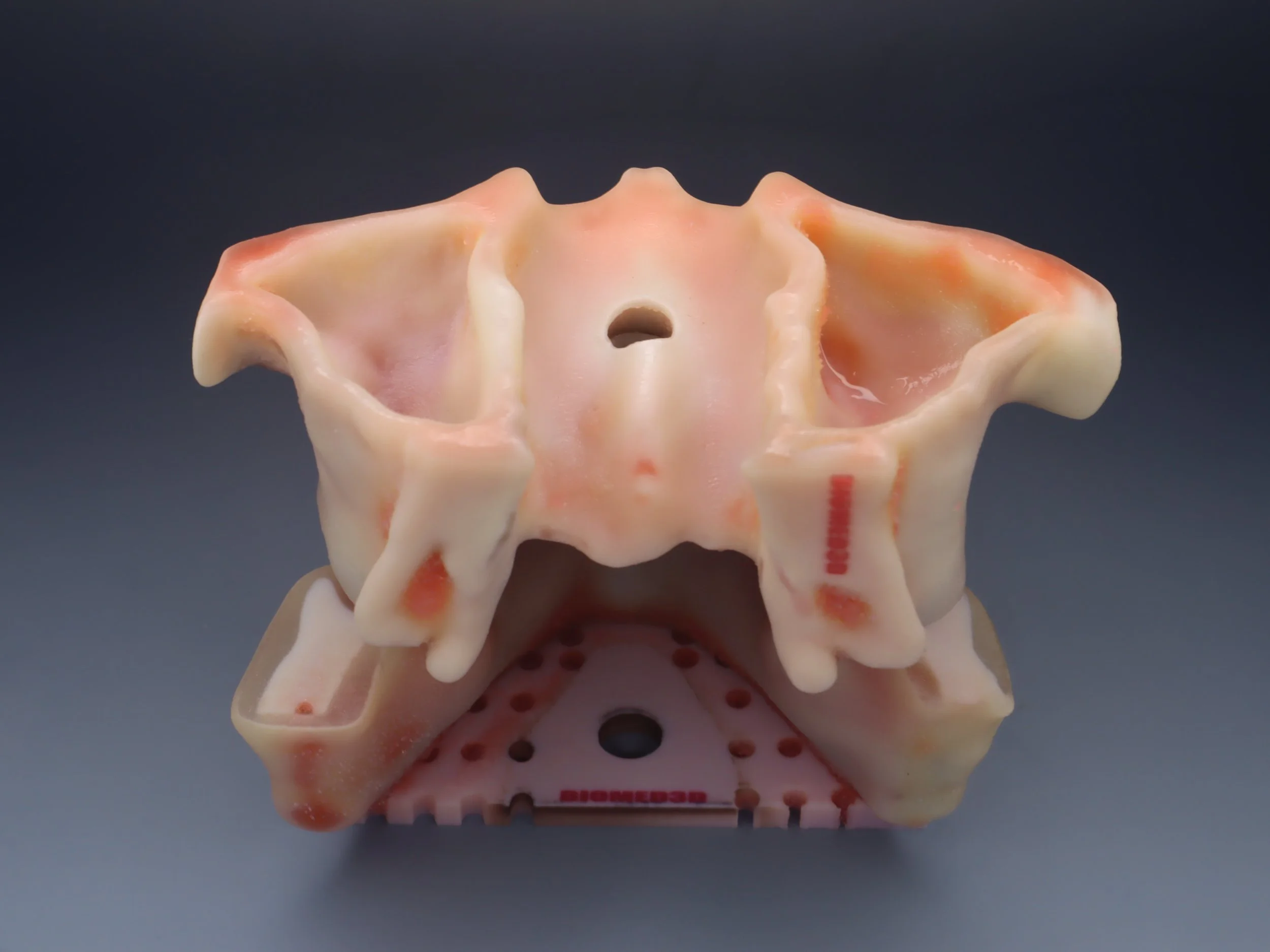
• Greater palatine groove and nerve landmarks
• Incisive canal region anatomy
• Sinus floor proximity simulation
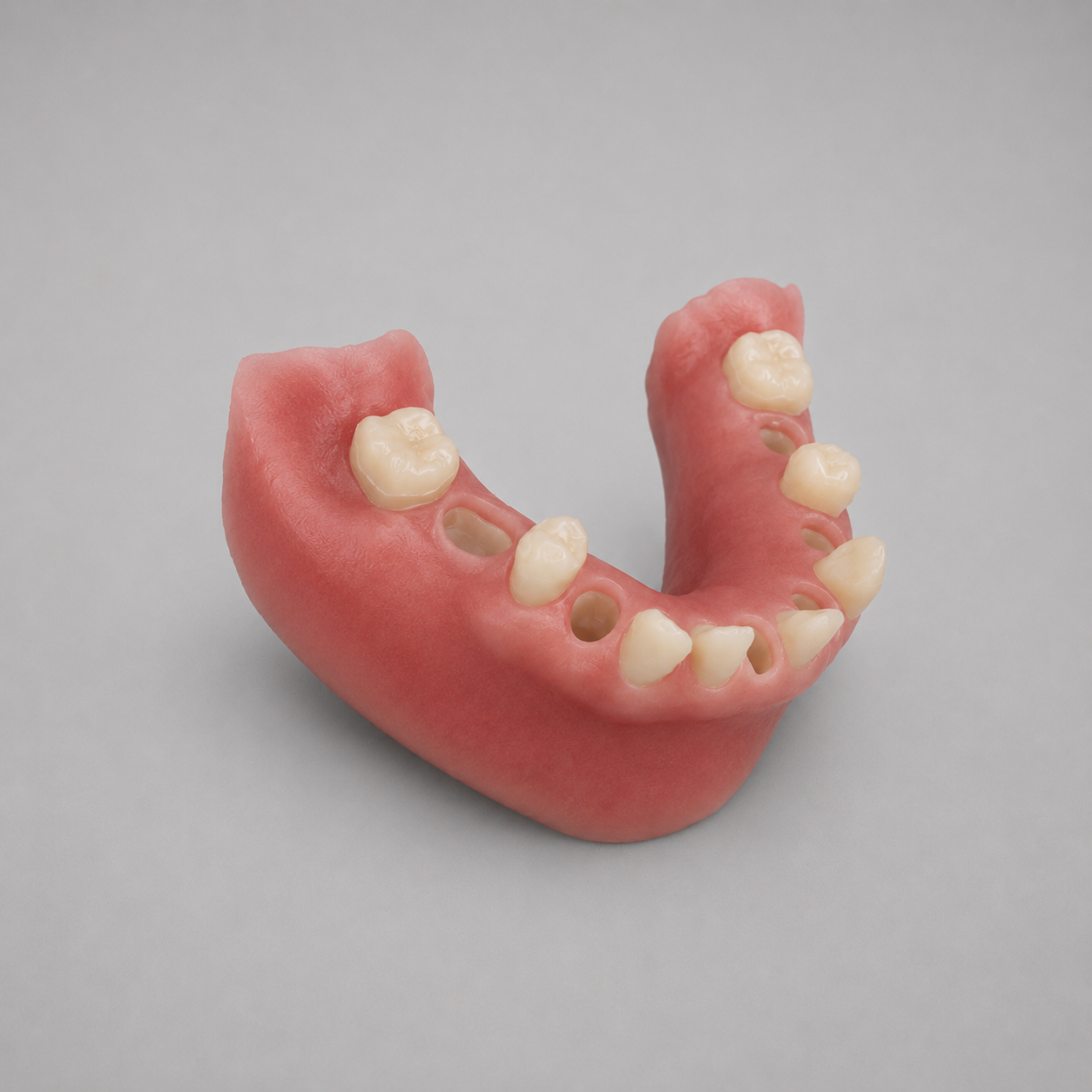
• Optional soft tissue for incision and suturing
• Radiographic compatible for x ray and CBCT review
• Durable premium training materials
Who Uses This
• Oral surgery programs
• Implant educators
• Dental schools
• Residency programs
• CE instructors
• Private training centers
3D-Viewer CLICK HERE
Train advanced maxillary oral surgery procedures with our Maxillary Extraction Model with Third Molars, Greater Palatine, and Incisive Canal Nerve Anatomy designed for dental schools, CE courses, OMS programs, implant educators, and clinicians.
This model simulates realistic maxillary anatomy for extractions, flap design, implant planning, sinus-related procedures, and anatomical landmark training.
Includes third molars, sinus proximity, palatal nerve landmarks, and soft tissue options for comprehensive hands-on education.
Best For
• Maxillary third molar extraction training
• Flap elevation and access design
• Sectioning and osteotomy practice
• Implant placement planning
• Sinus proximity case training
• Suturing techniques
• Palatal anatomy education
Key Features
• Realistic maxillary jaw anatomy
• Includes upper third molars
• Greater palatine groove and nerve landmarks
• Incisive canal region anatomy
• Sinus floor proximity simulation
• Optional soft tissue for incision and suturing
• Radiographic compatible for x ray and CBCT review
• Durable premium training materials
Who Uses This
• Oral surgery programs
• Implant educators
• Dental schools
• Residency programs
• CE instructors
• Private training centers

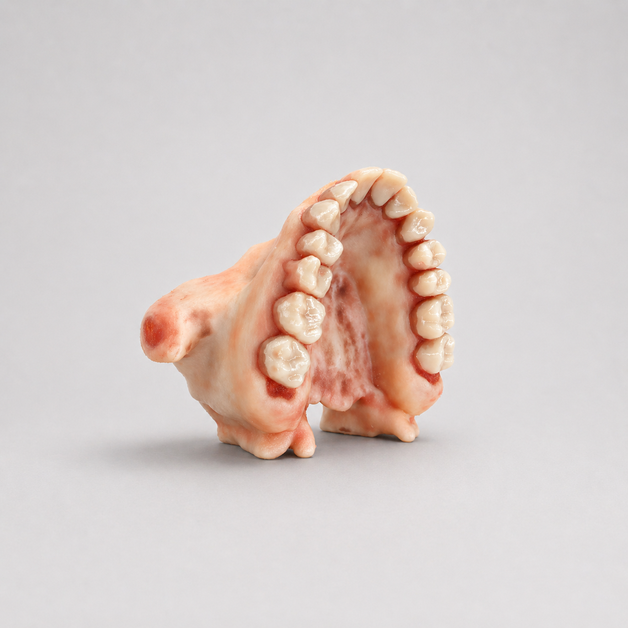 Image 1 of 22
 Image 2 of 22
 Image 3 of 22
 Image 4 of 22
 Image 5 of 22
 Image 6 of 22
 Image 7 of 22
 Image 8 of 22
 Image 9 of 22
 Image 10 of 22
 Image 11 of 22
 Image 12 of 22
 Image 13 of 22
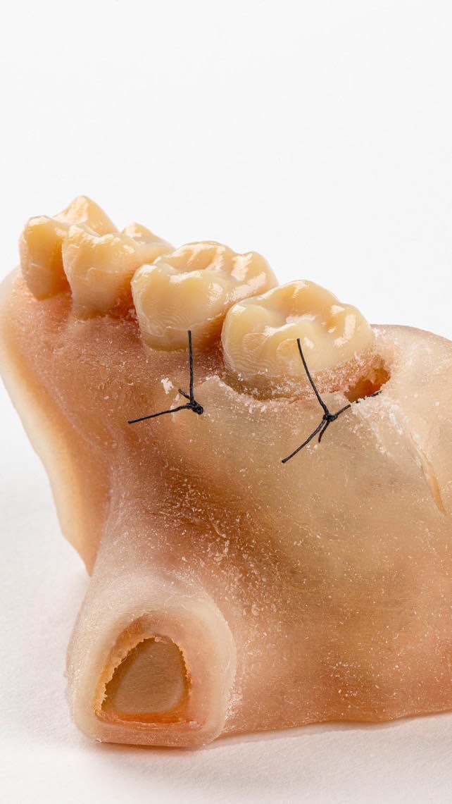 Image 14 of 22
 Image 15 of 22
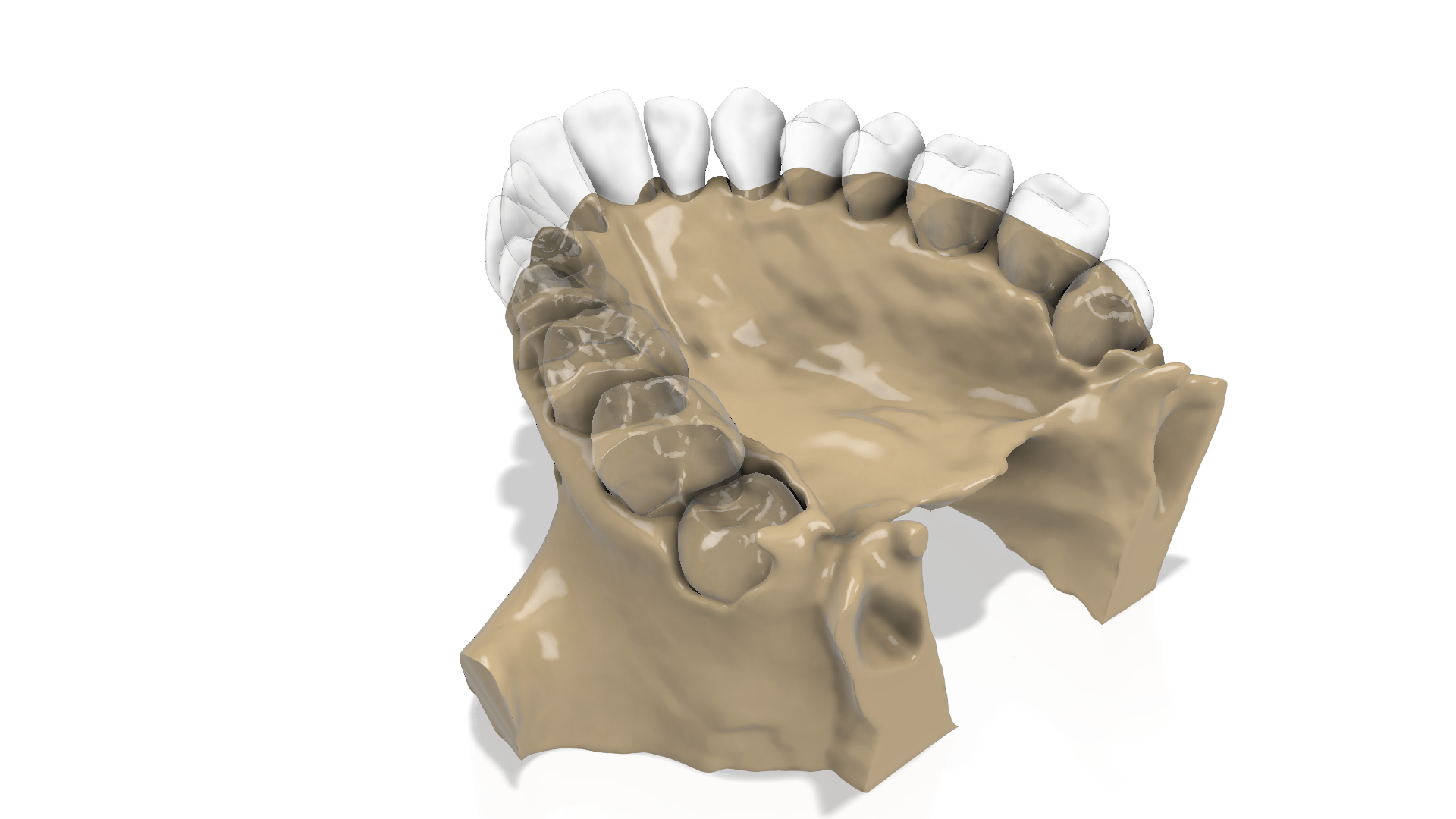 Image 16 of 22
 Image 17 of 22
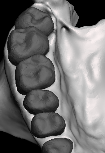 Image 18 of 22
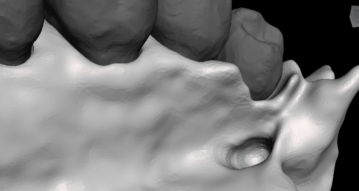 Image 19 of 22
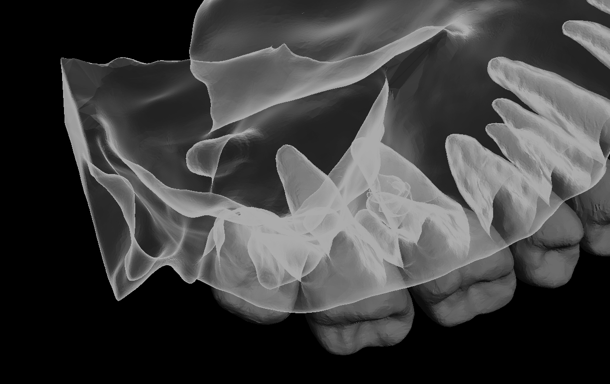 Image 20 of 22
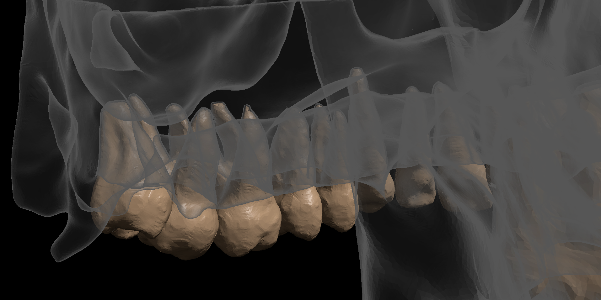 Image 21 of 22
 Image 22 of 22