 Image 1 of 5
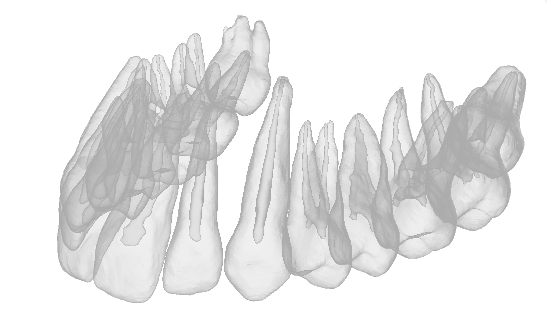 Image 2 of 5
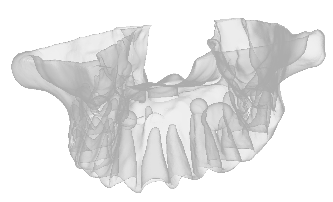 Image 3 of 5
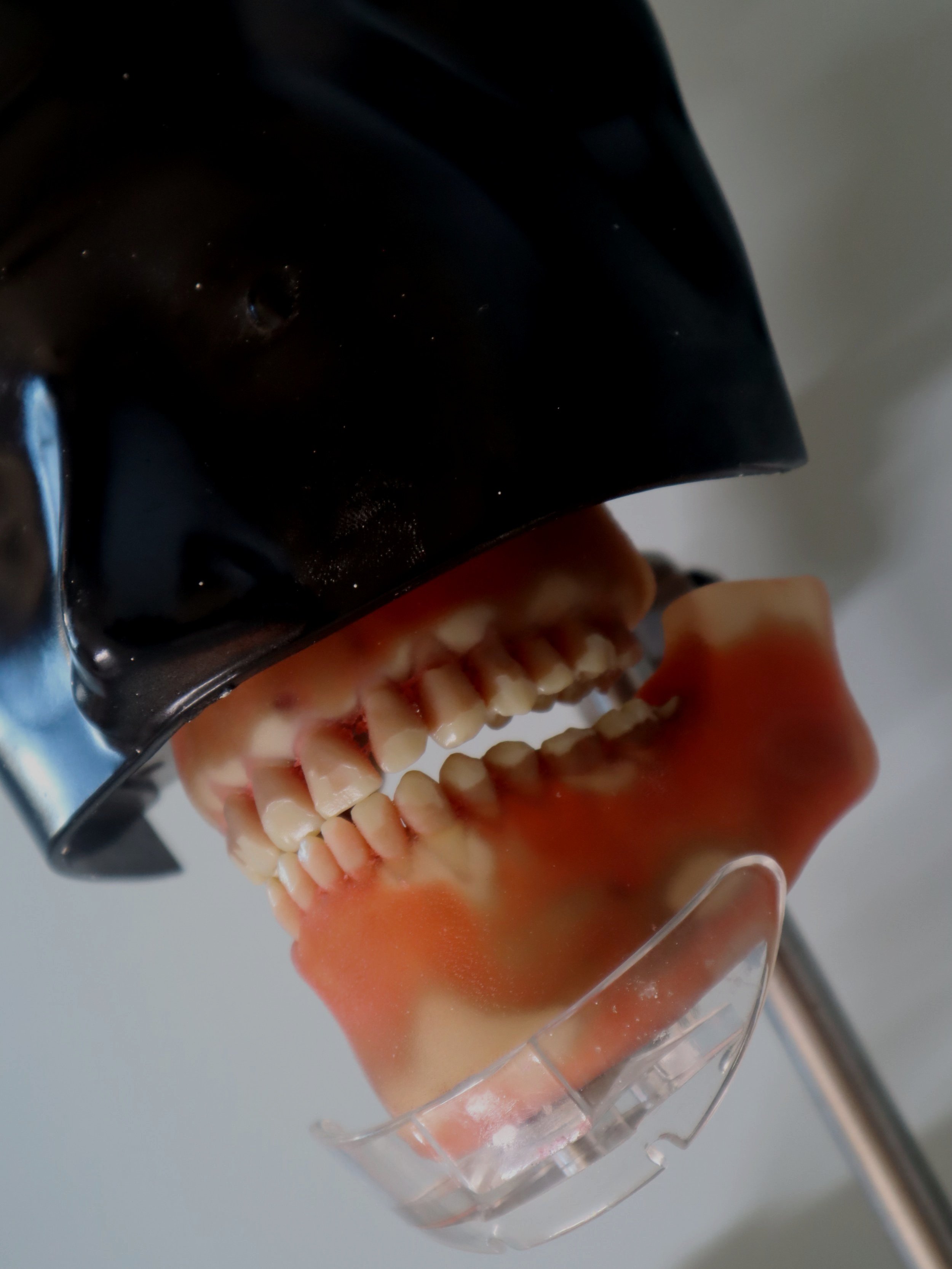 Image 4 of 5
 Image 5 of 5
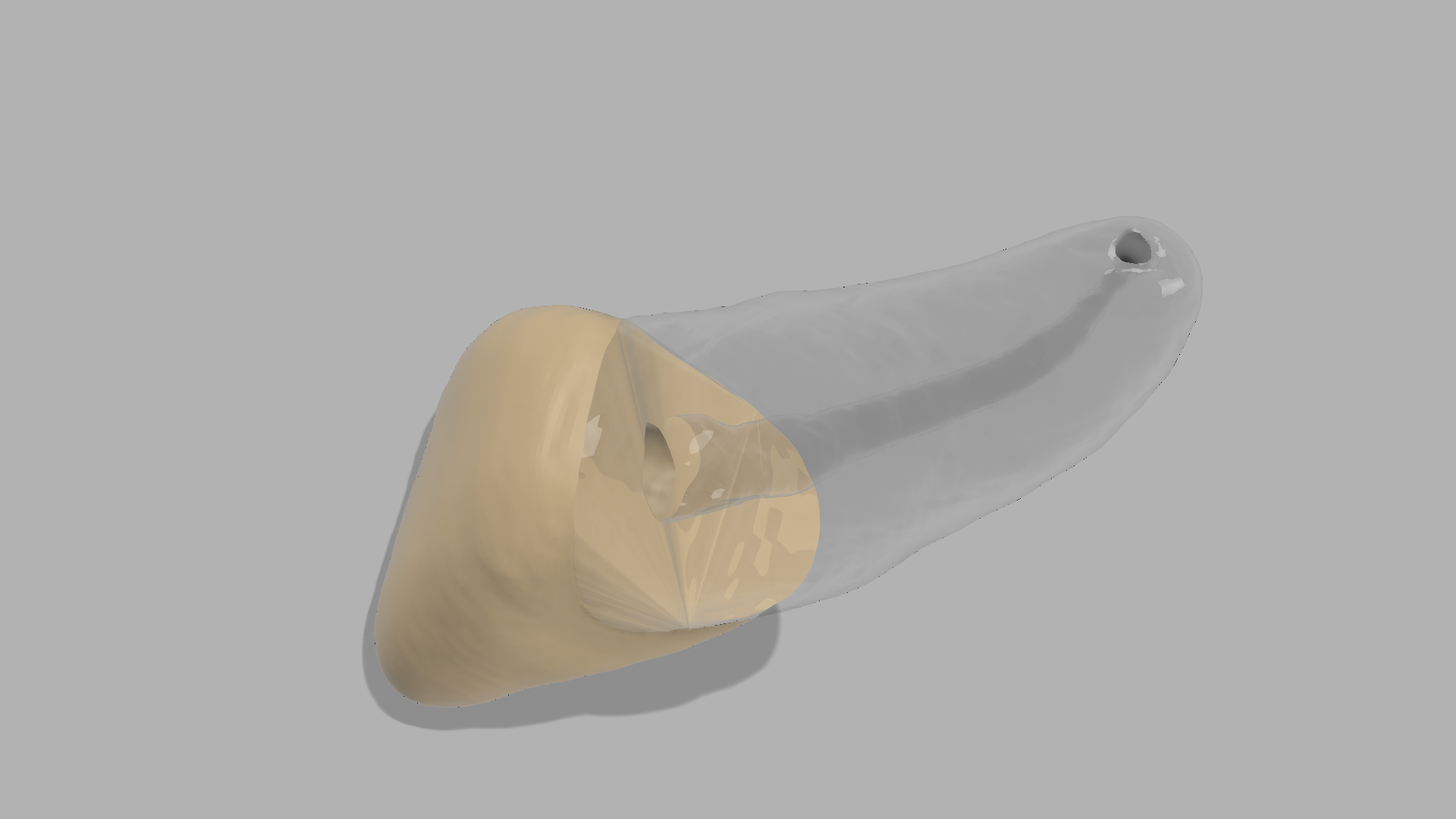
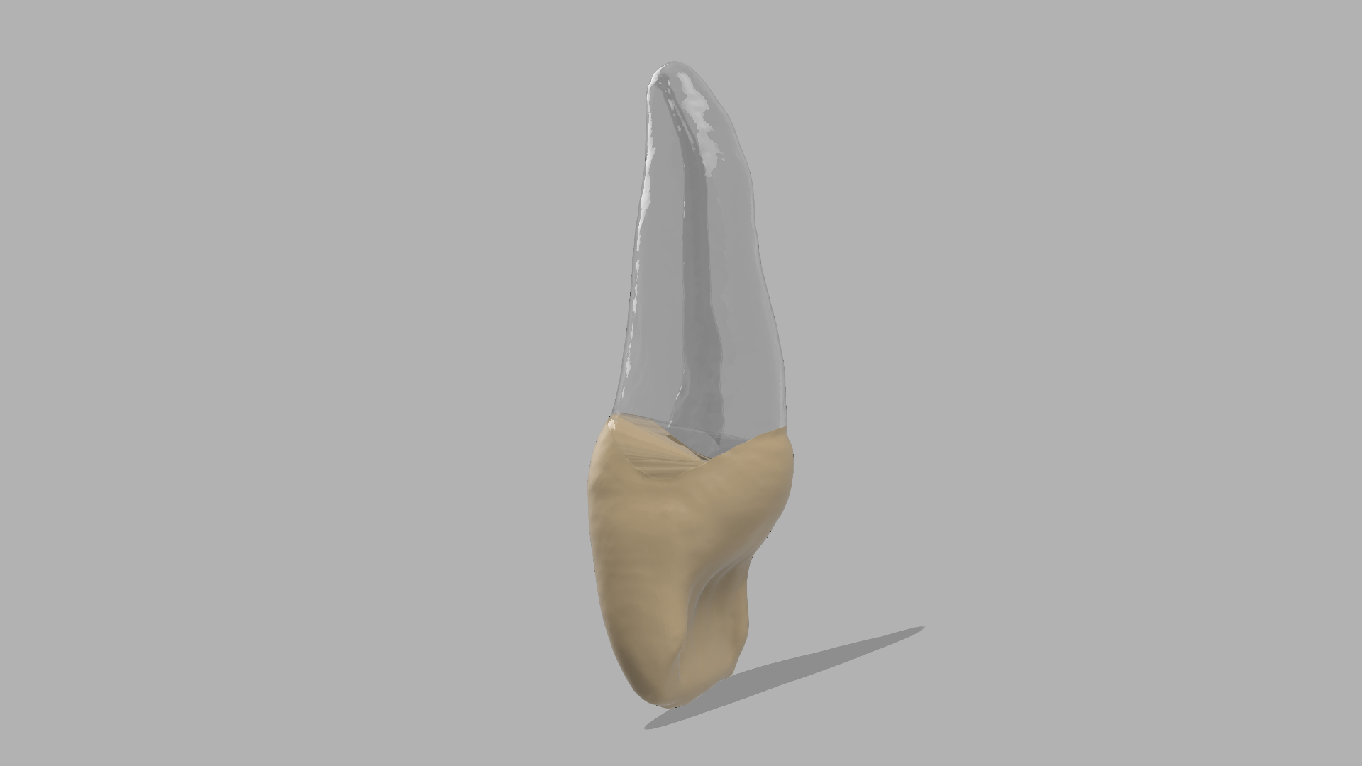
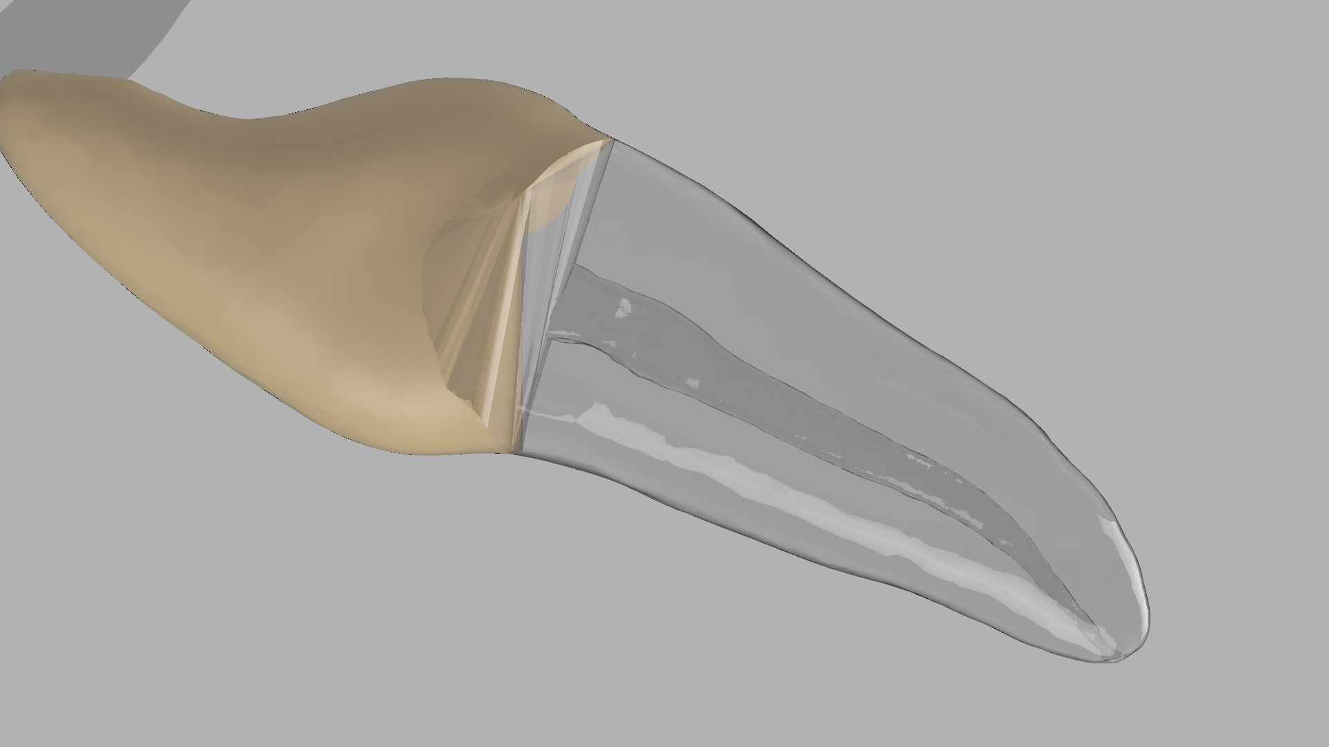
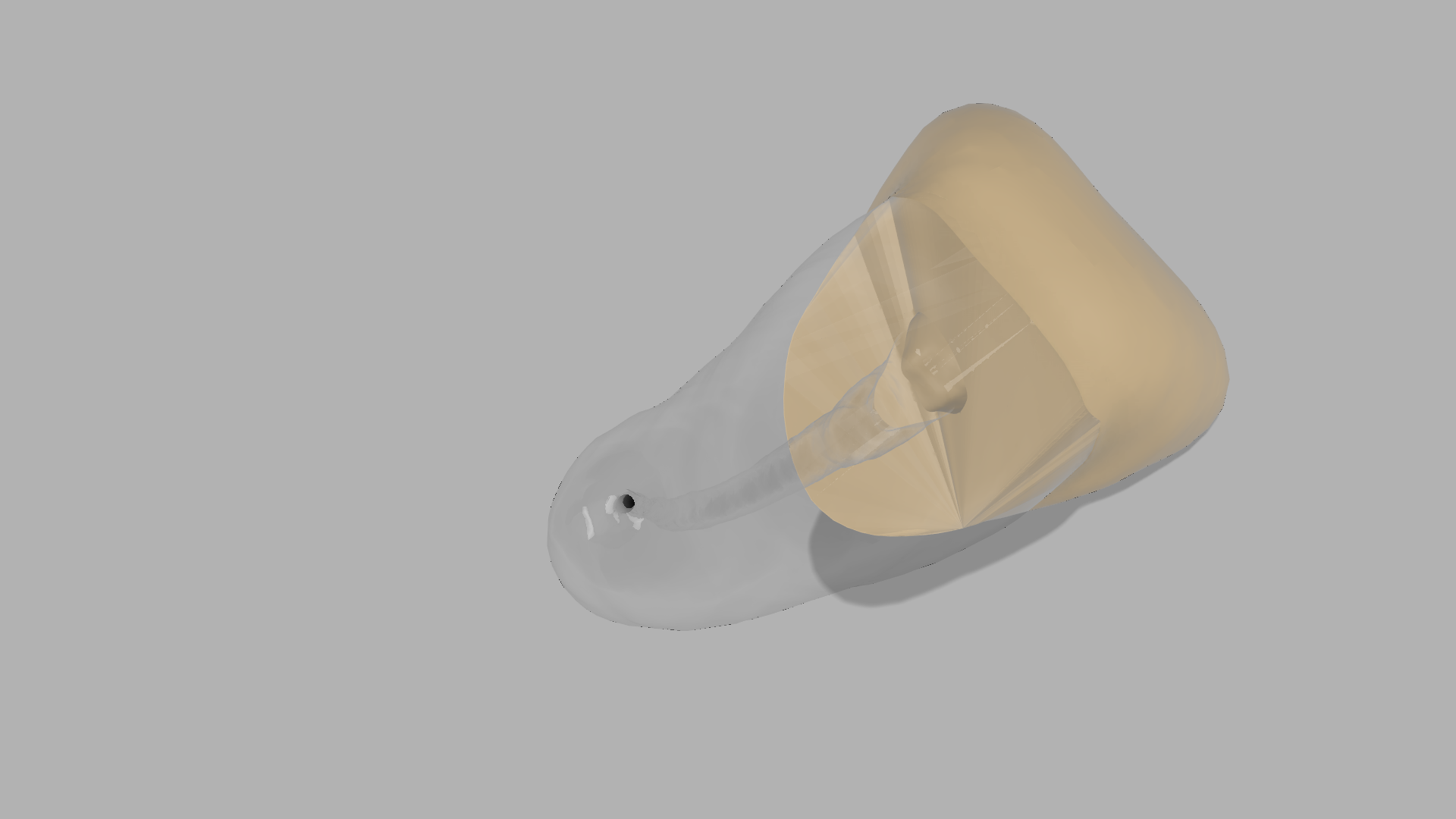
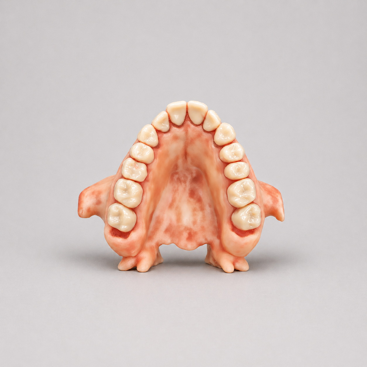
Train advanced endodontic microsurgery with our BIOJAW Maxillary Endodontic Apical Surgery Model designed for endodontists, oral surgeons, residency programs, and CE educators.
This model simulates multiple maxillary apical surgery cases in one arch, allowing hands-on training for flap design, osteotomy, lesion curettage, root-end resection, retroprep, grafting, and suturing.
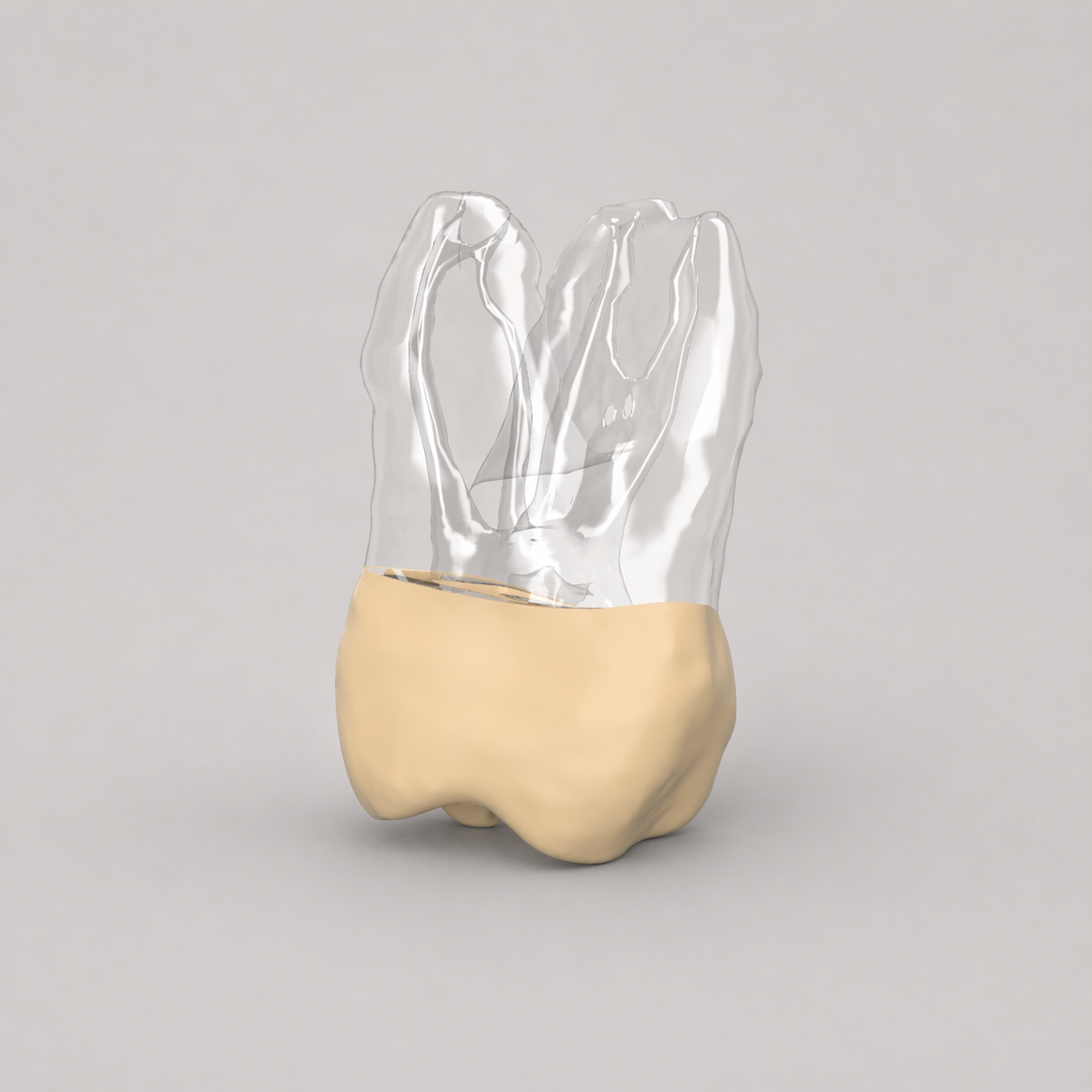
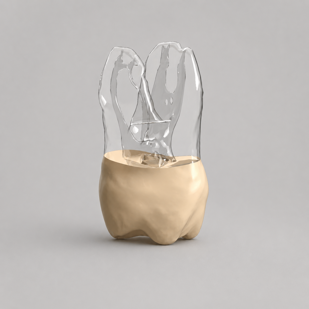
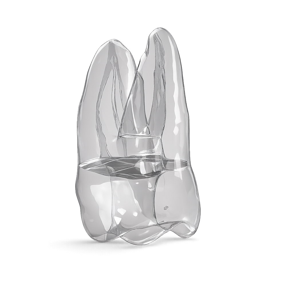
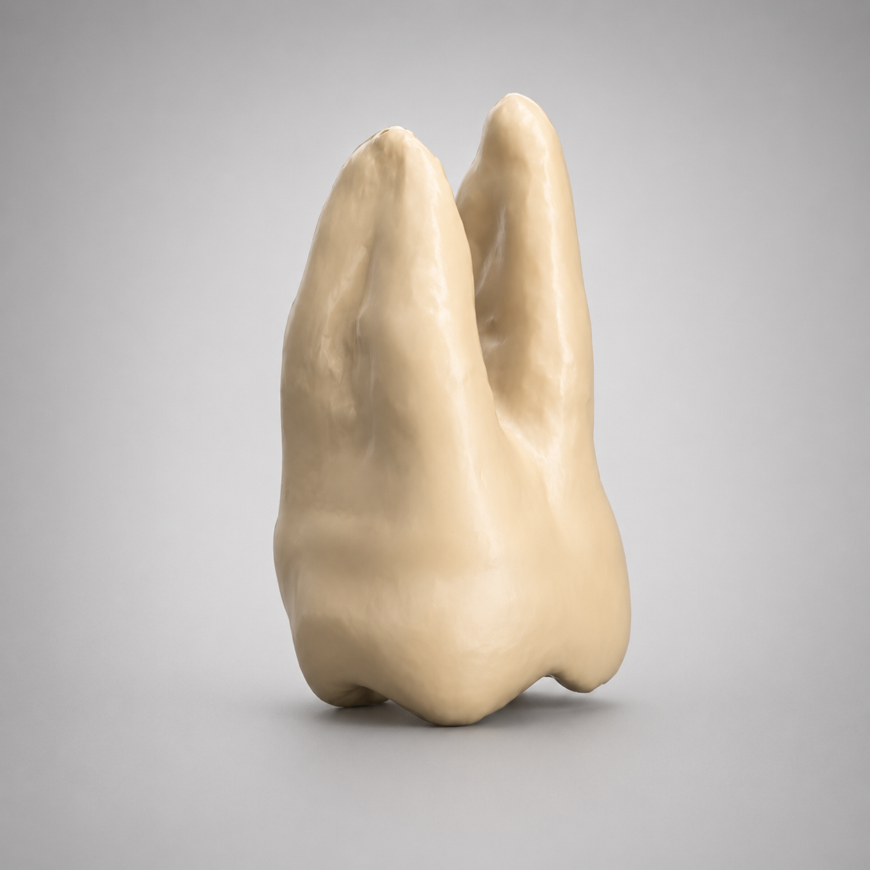
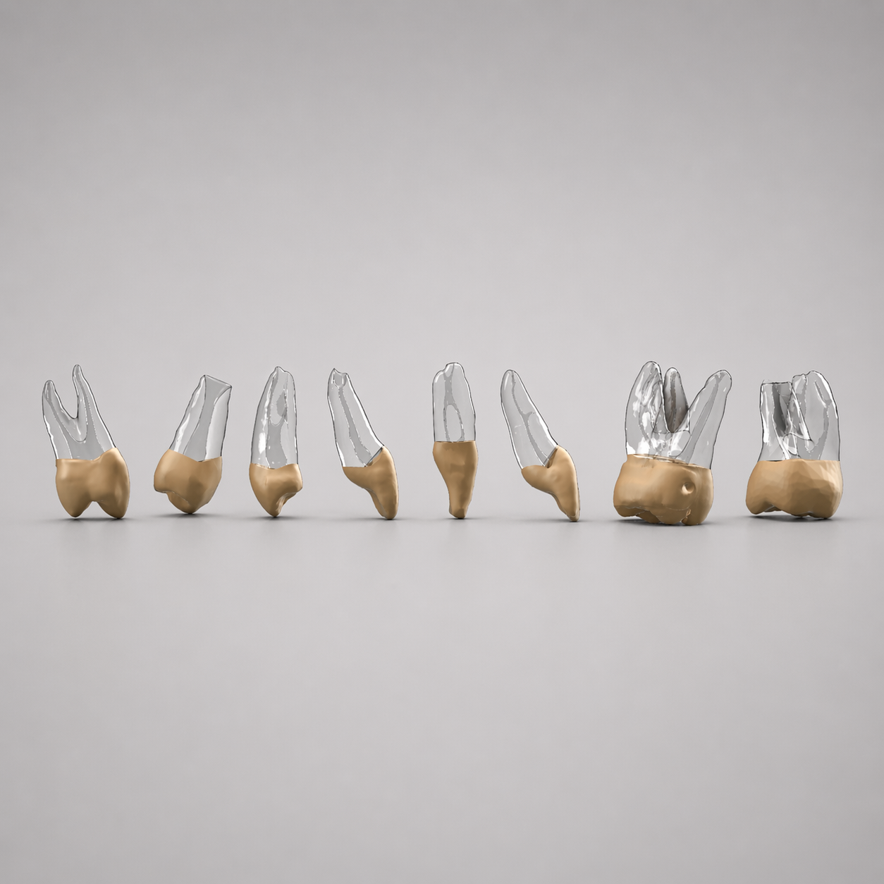
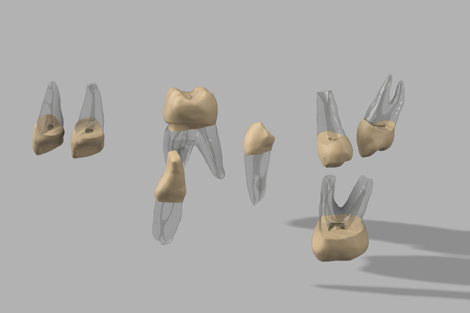
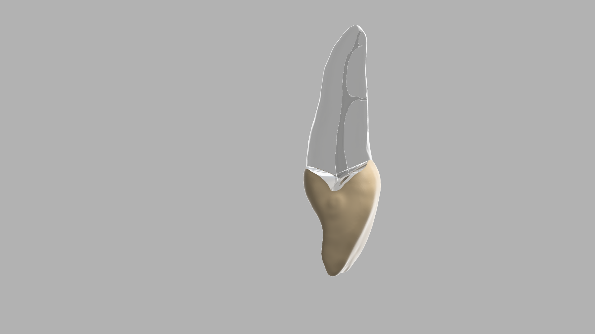
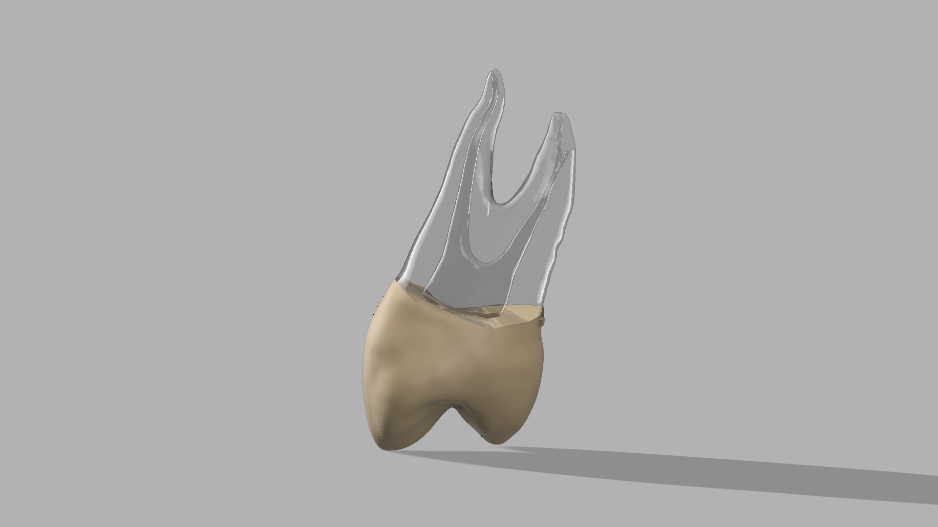
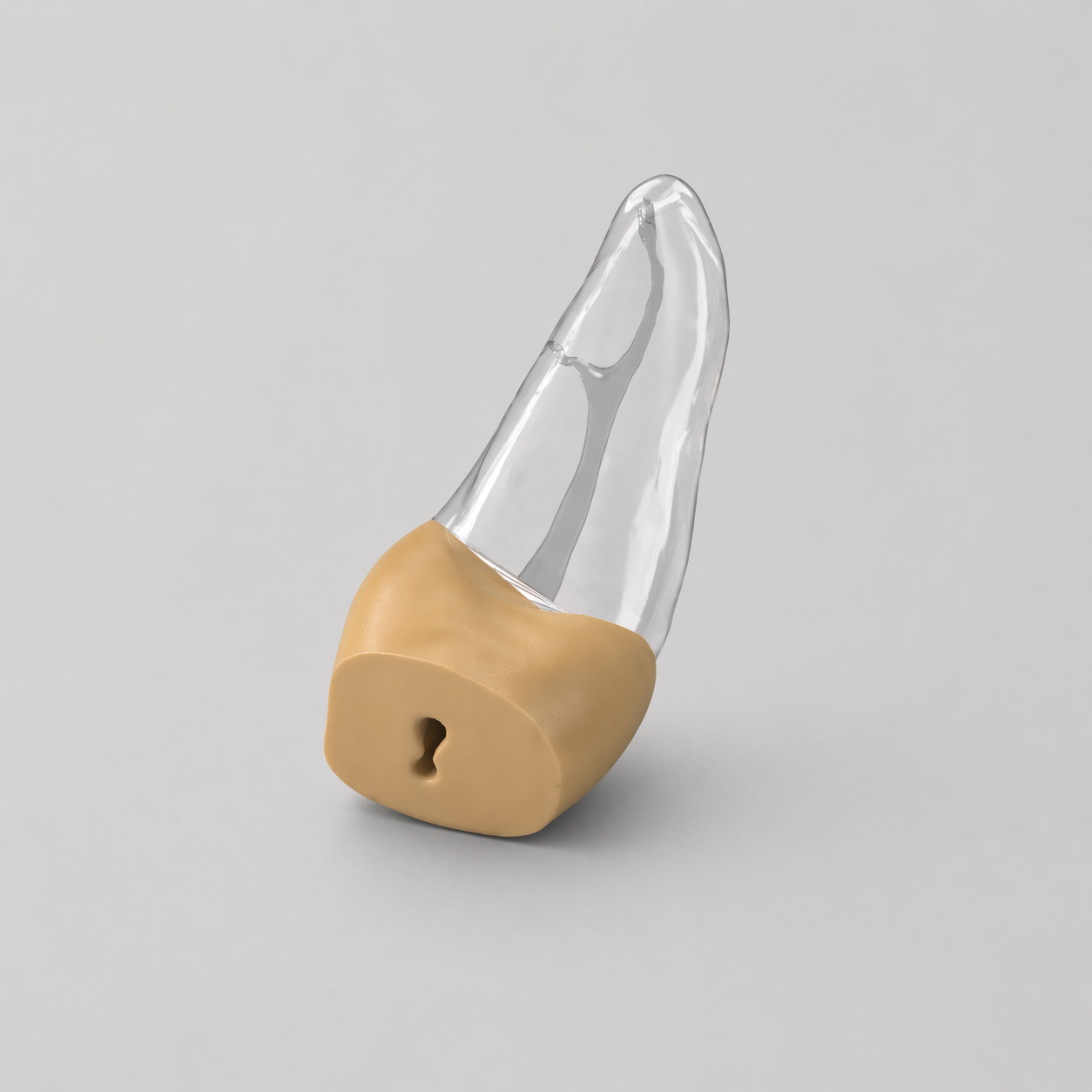
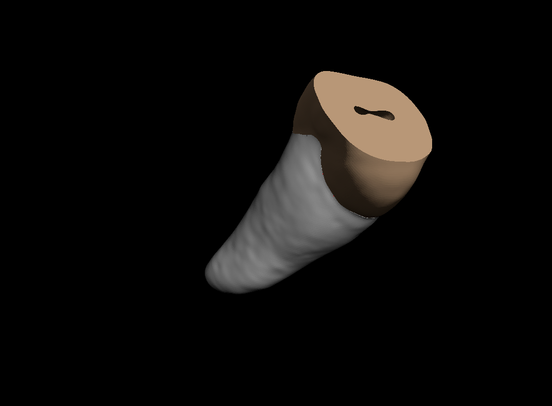
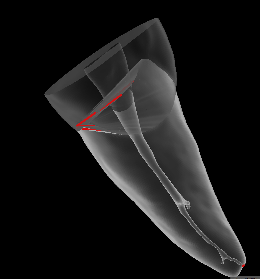
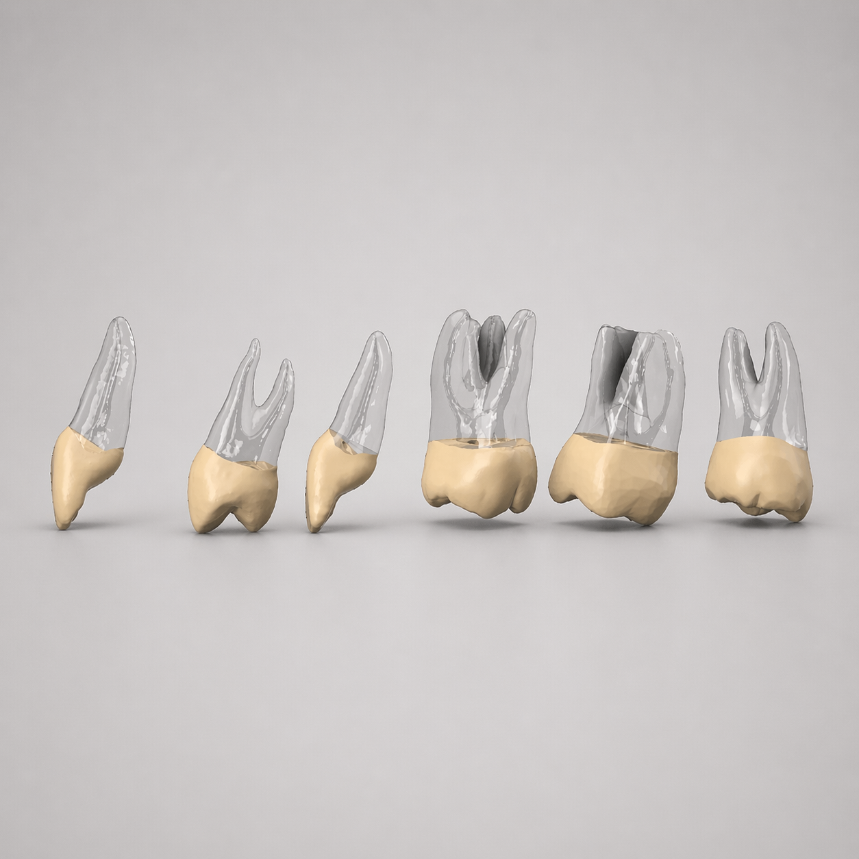
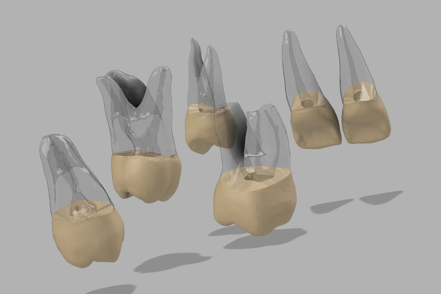
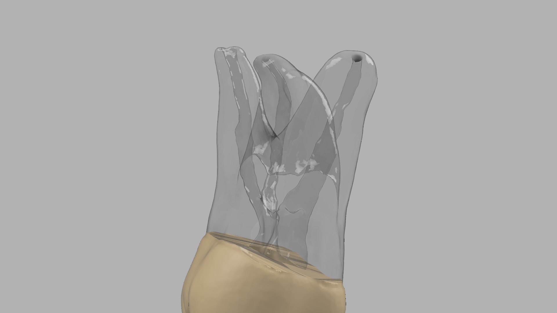
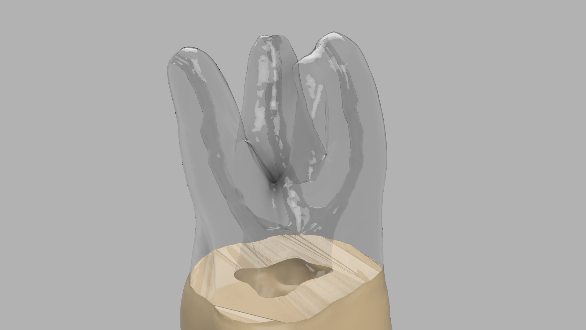
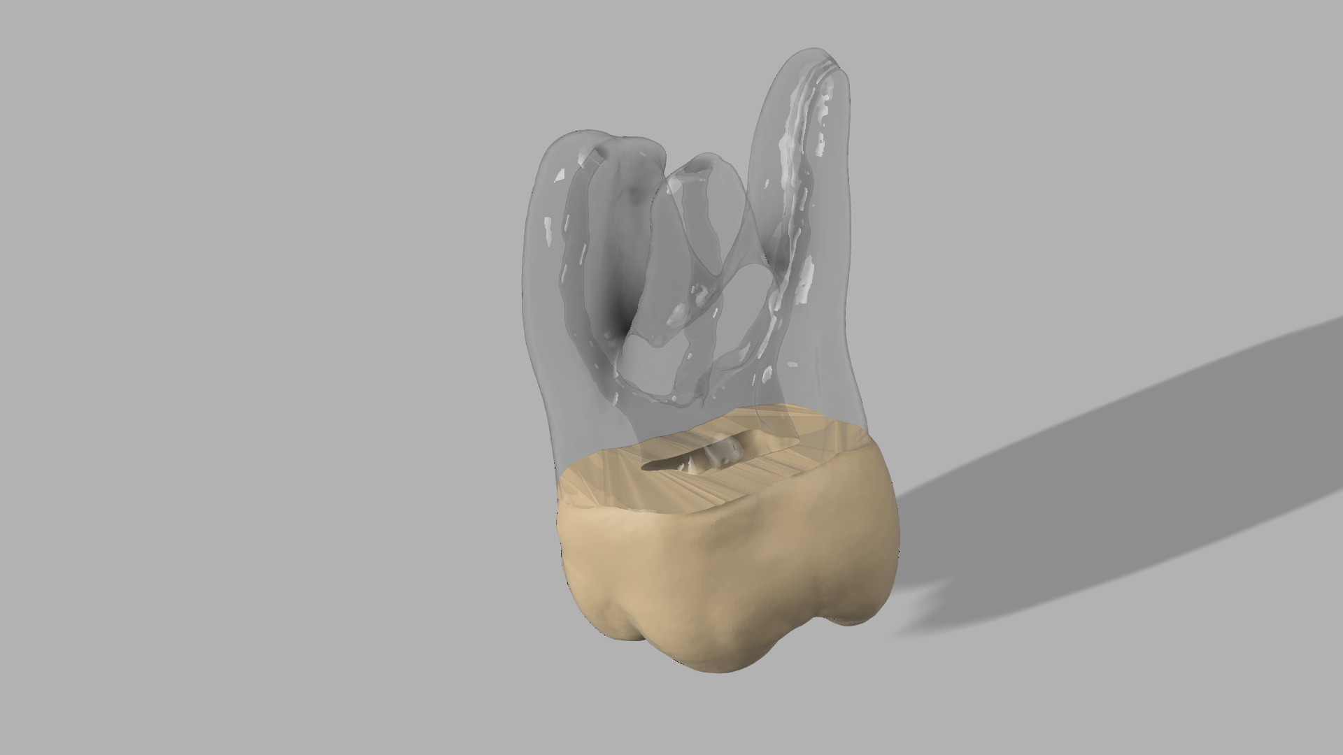
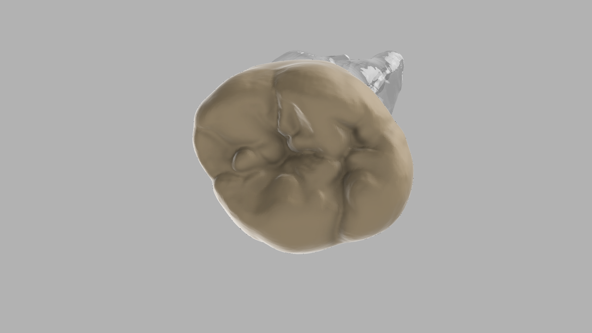
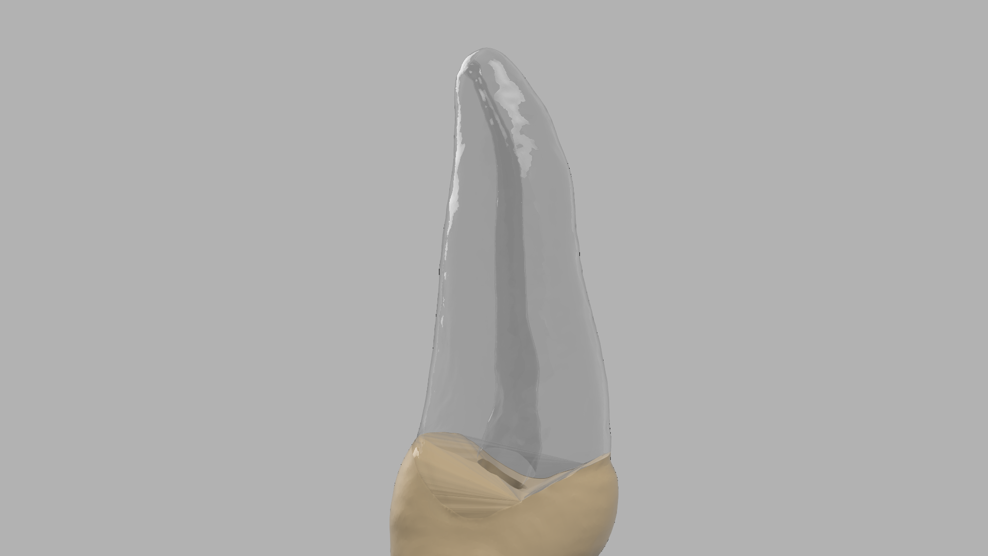
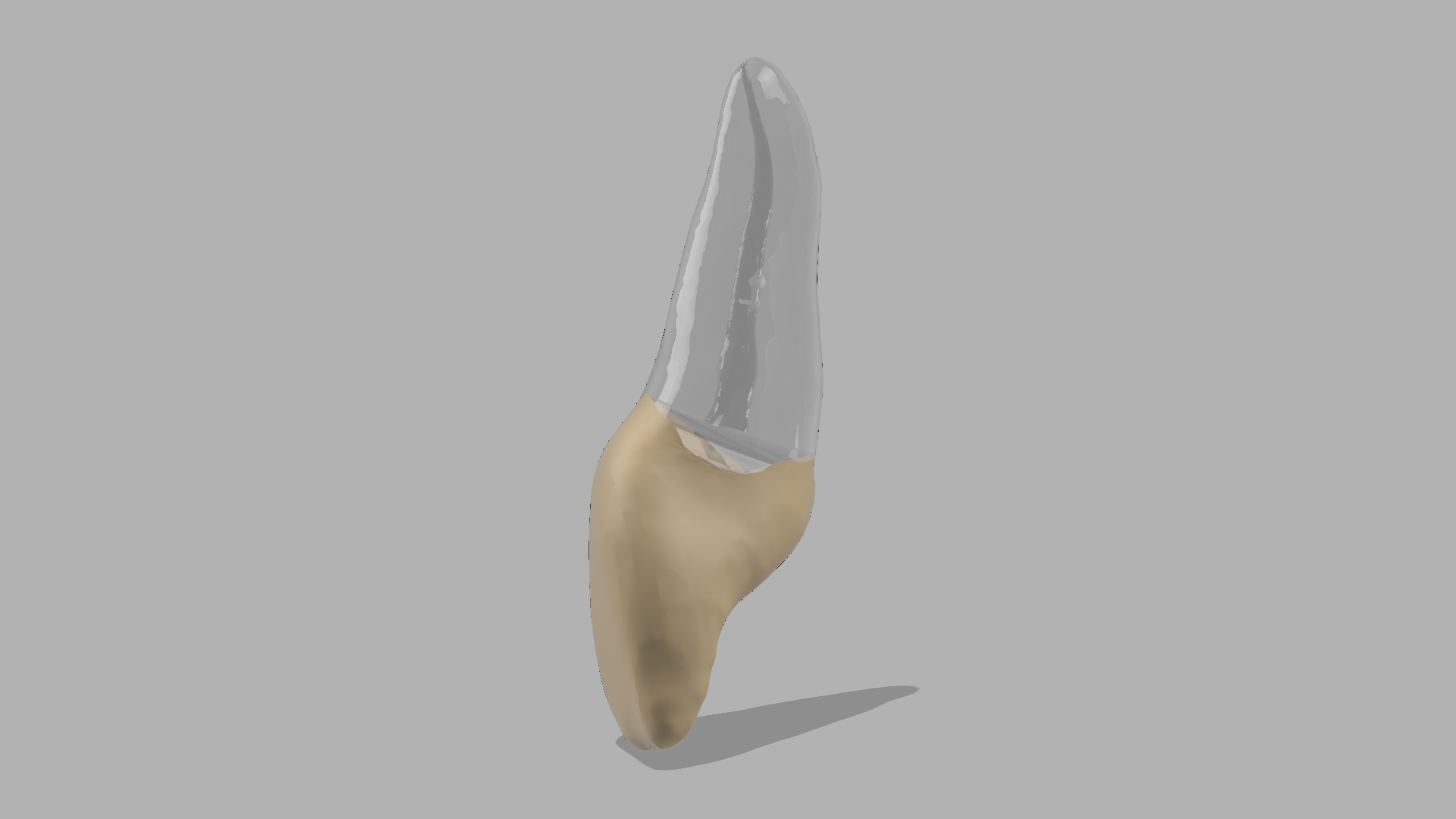
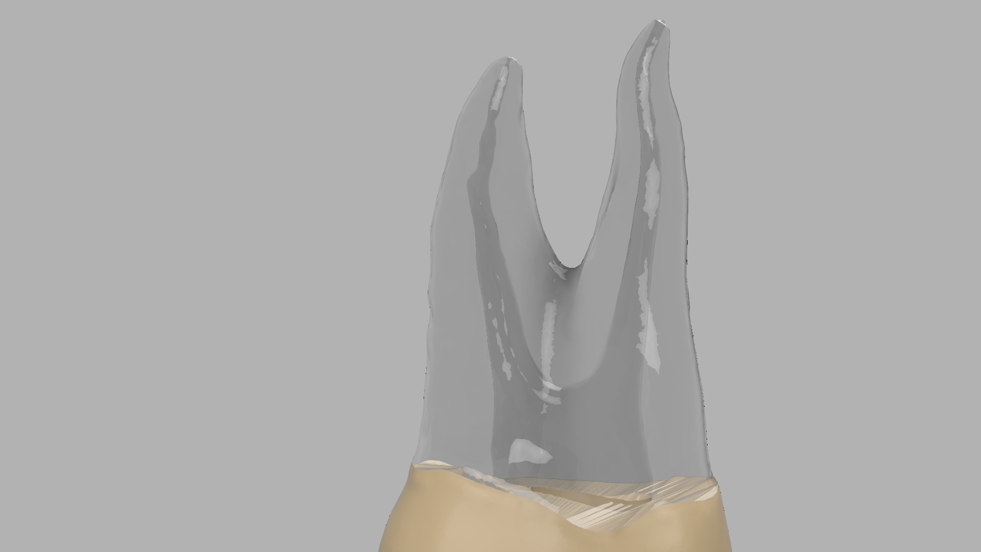
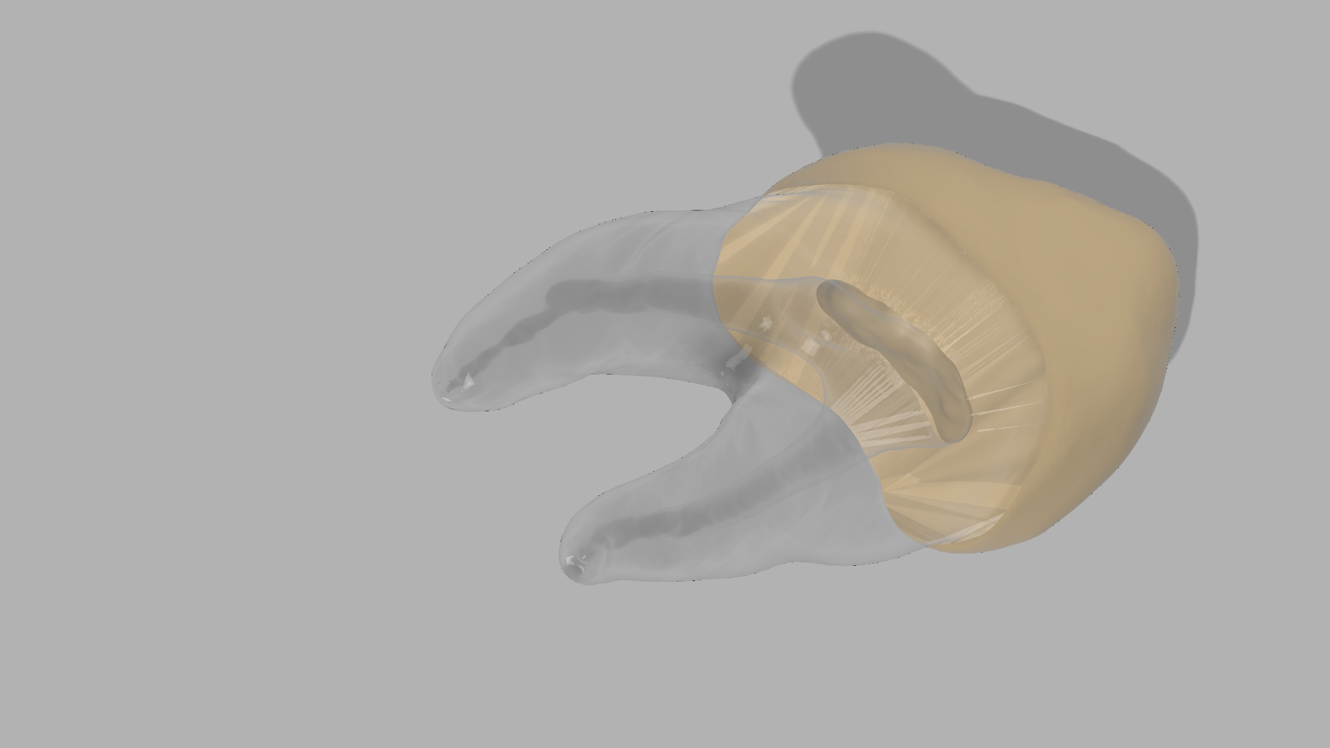
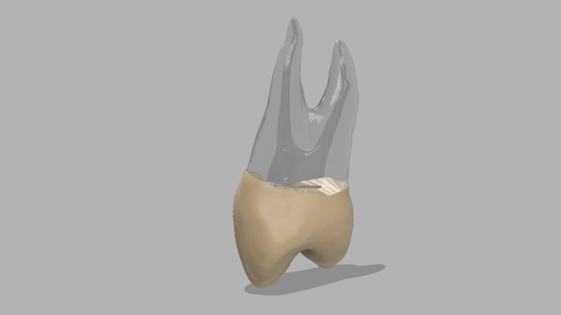
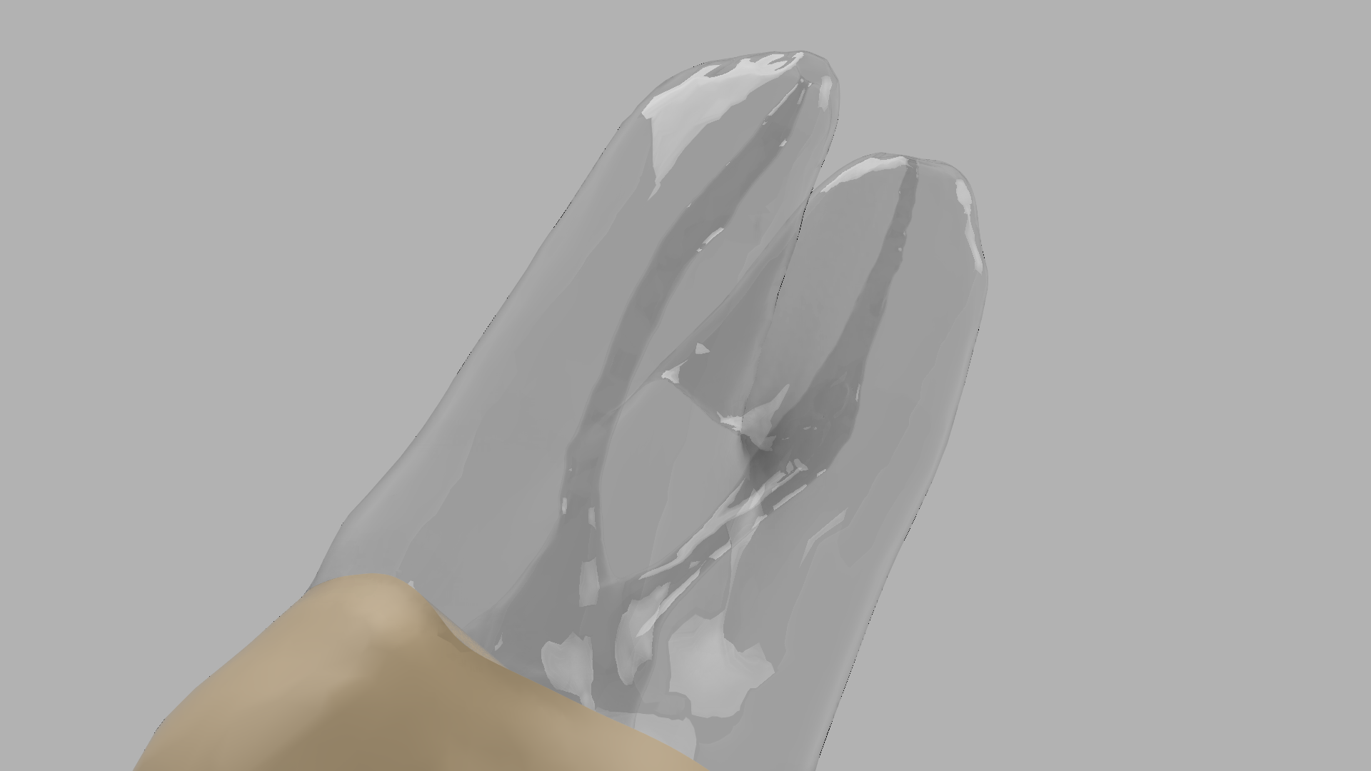
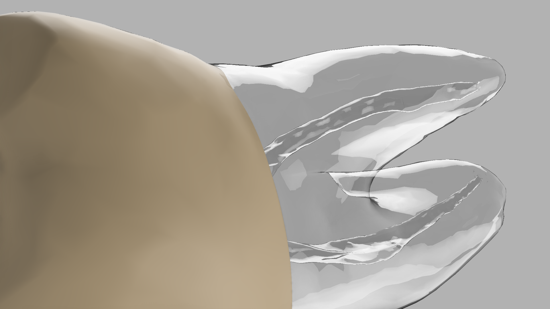
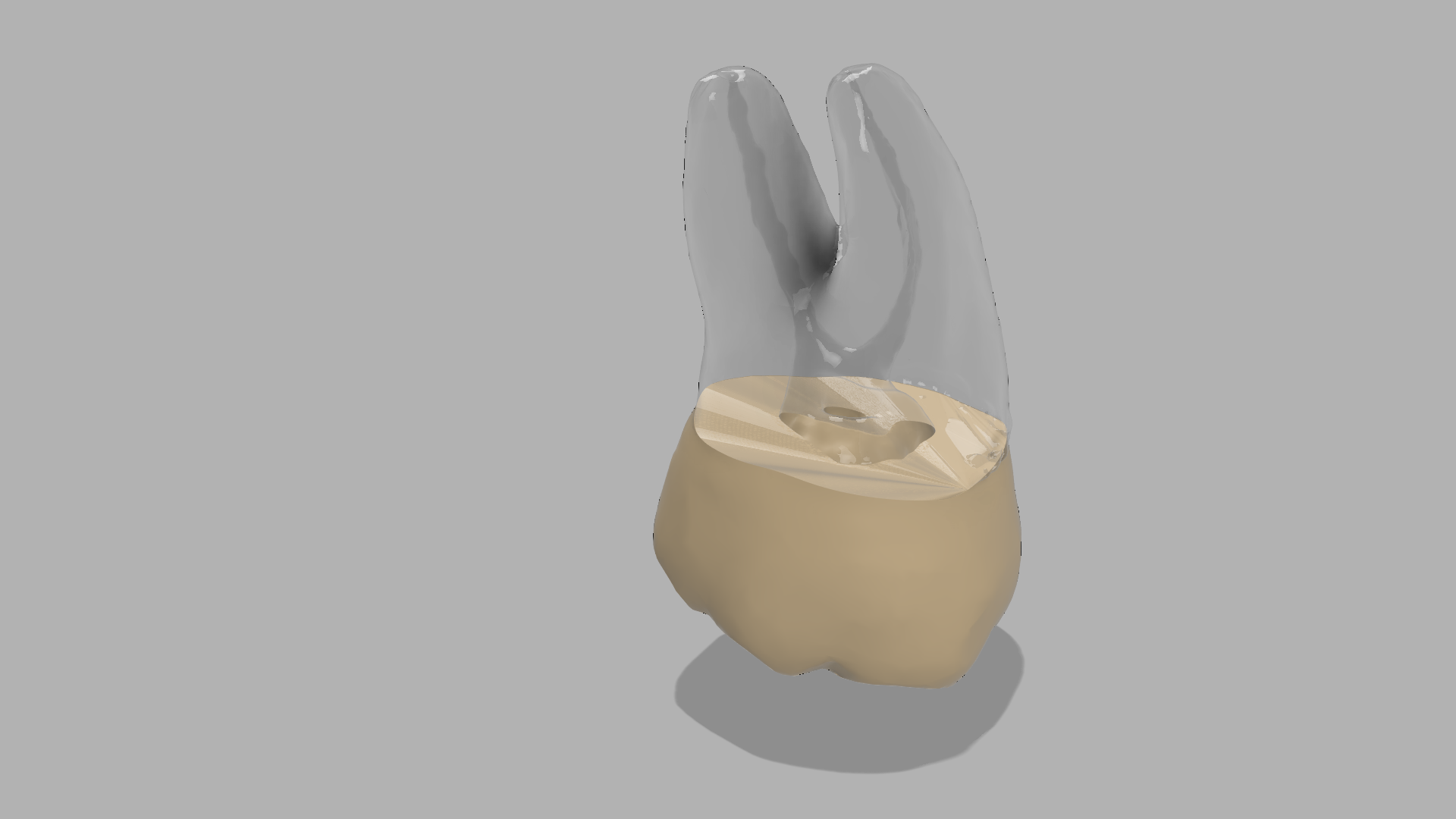
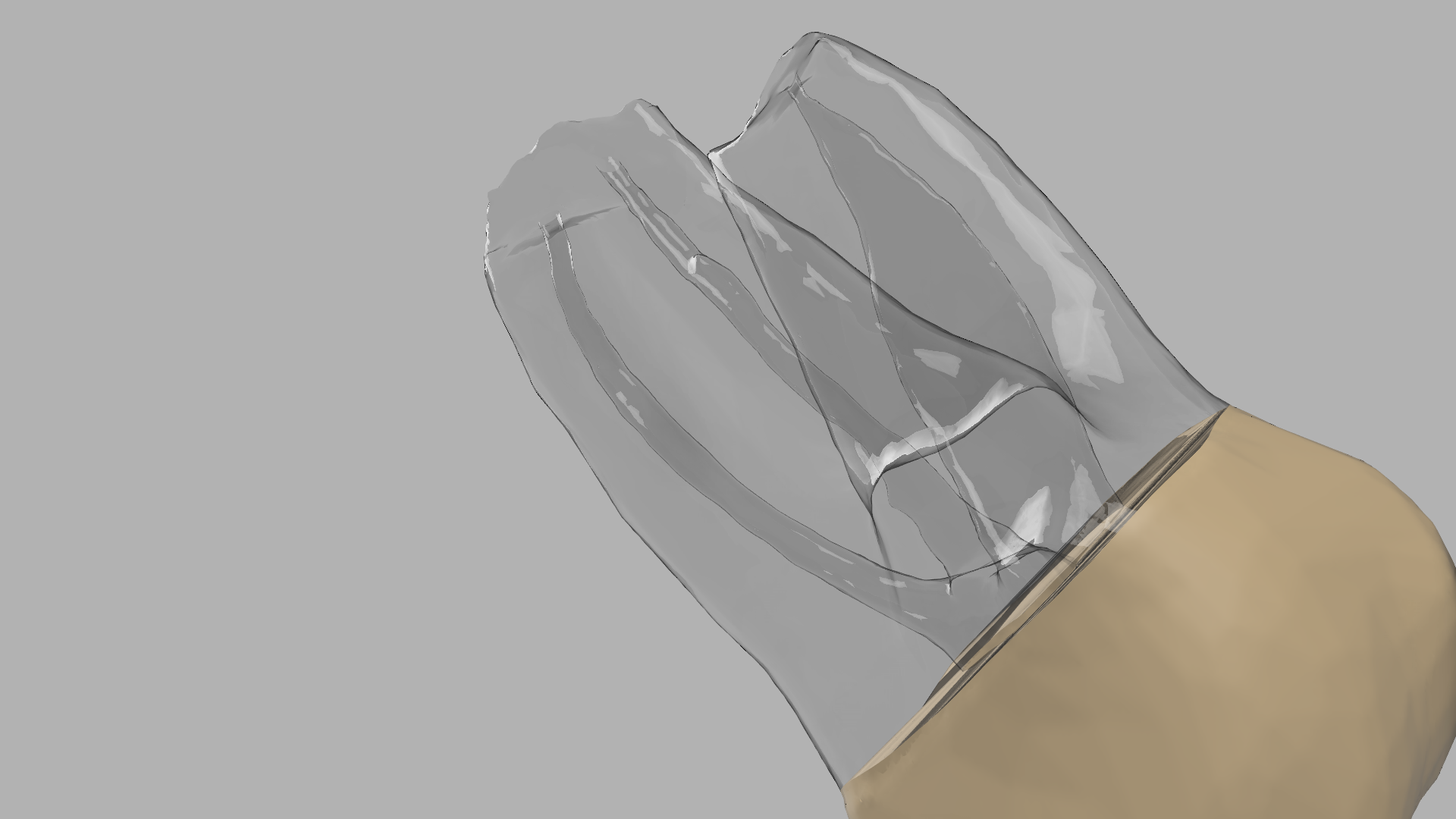
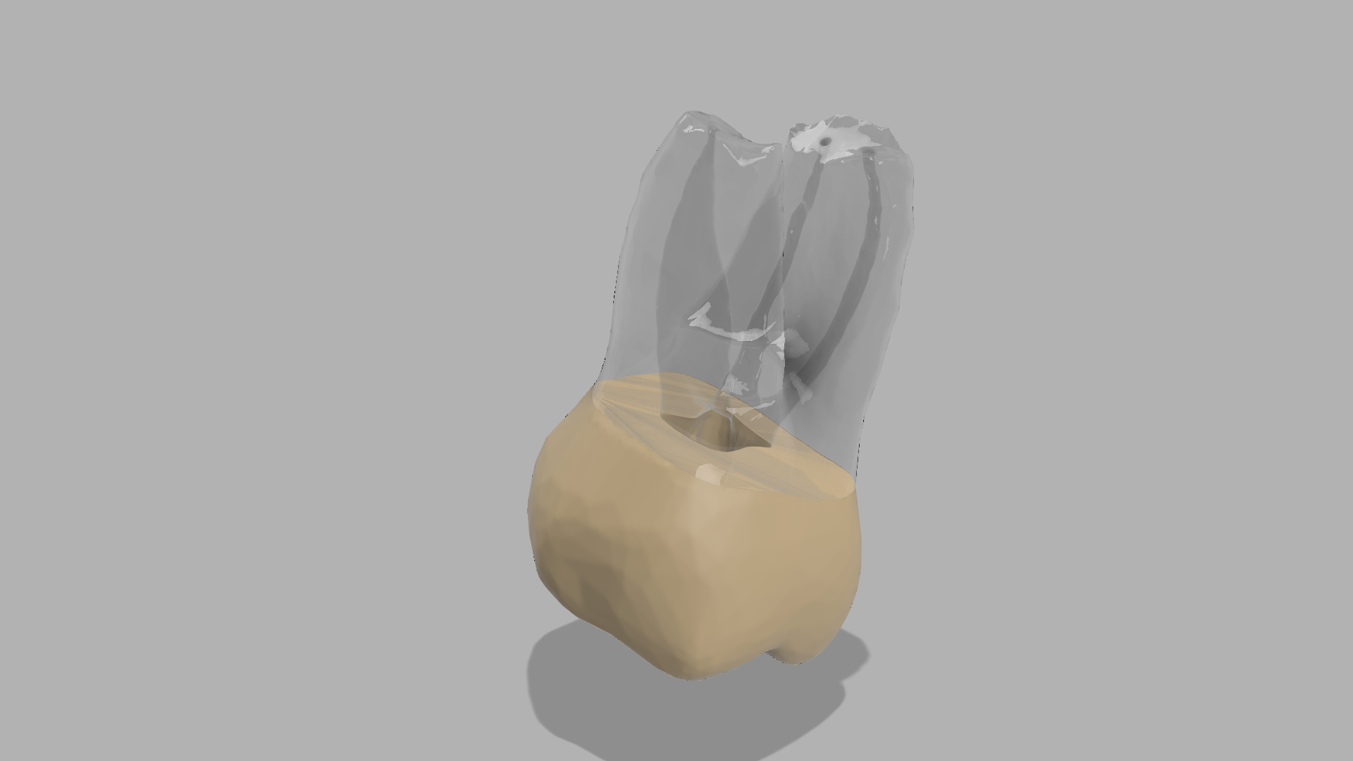
Built from real anatomical data with internal root anatomy, cortical and cancellous bone representation, and radiopaque material for CBCT and x ray planning.
• Tooth #2 posterior maxillary apicoectomy case
• Tooth #6 canine apical lesion case
• Tooth #8 anterior central incisor lesion case
• Teeth #11, #12, #13 multi-tooth confluent lesion case
• Tooth #14 multi-root posterior lesion case
• Apicoectomy training
• Root-end resection practice
• Retroprep and retrofill training
• Lesion curettage procedures
• Flap elevation and suturing
• Bone graft decision-making
• Microsurgical CE workshops
• Multiple surgical cases in one maxillary model
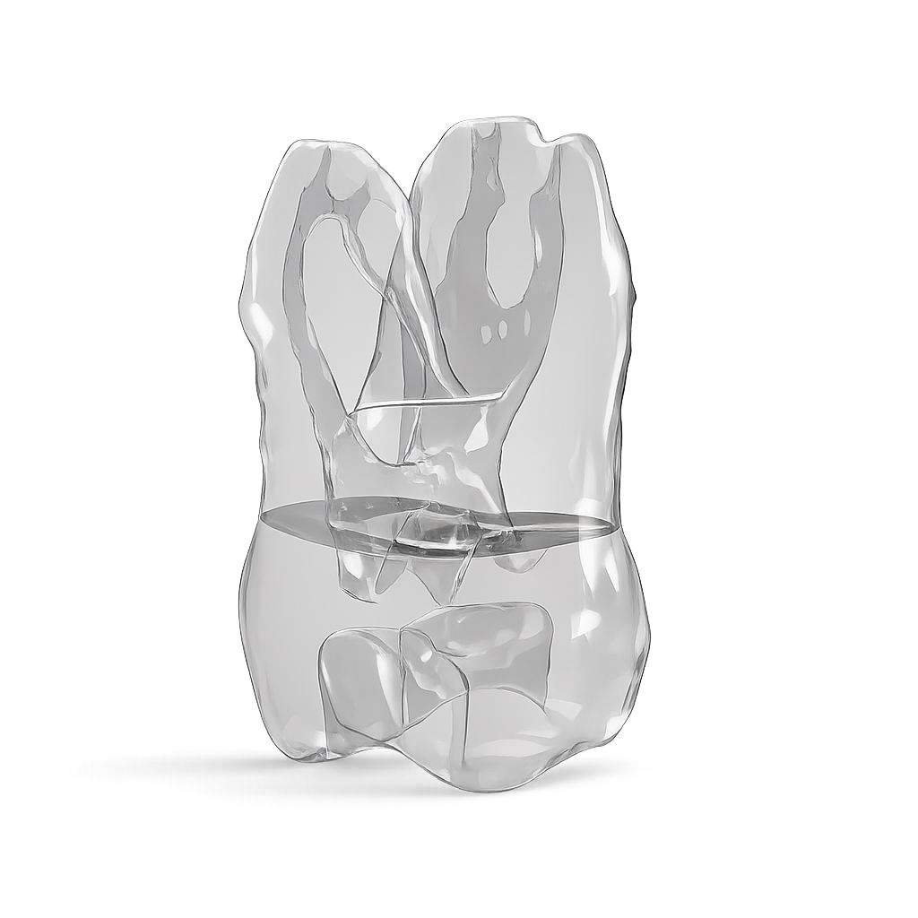
• Internal root anatomy included
• Cortical and cancellous bone simulation
• Soft tissue layer for incision and suturing
• Radiopaque for CBCT and x ray checks
• Handheld or phantom head compatible
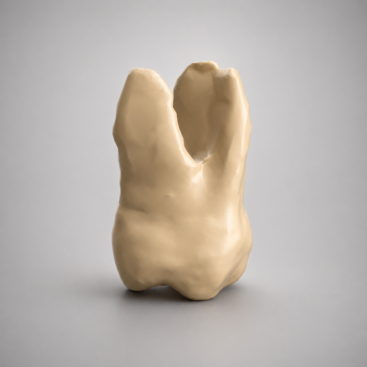
• Available in opaque or transparent variants
• Durable premium training materials
• Endodontic residency programs
• Oral surgery programs
• CE educators
• Dental schools
• Specialists
• Private training centers
Train advanced endodontic microsurgery with our BIOJAW Maxillary Endodontic Apical Surgery Model designed for endodontists, oral surgeons, residency programs, and CE educators.
This model simulates multiple maxillary apical surgery cases in one arch, allowing hands-on training for flap design, osteotomy, lesion curettage, root-end resection, retroprep, grafting, and suturing.
Built from real anatomical data with internal root anatomy, cortical and cancellous bone representation, and radiopaque material for CBCT and x ray planning.
• Tooth #2 posterior maxillary apicoectomy case
• Tooth #6 canine apical lesion case
• Tooth #8 anterior central incisor lesion case
• Teeth #11, #12, #13 multi-tooth confluent lesion case
• Tooth #14 multi-root posterior lesion case
• Apicoectomy training
• Root-end resection practice
• Retroprep and retrofill training
• Lesion curettage procedures
• Flap elevation and suturing
• Bone graft decision-making
• Microsurgical CE workshops
• Multiple surgical cases in one maxillary model
• Internal root anatomy included
• Cortical and cancellous bone simulation
• Soft tissue layer for incision and suturing
• Radiopaque for CBCT and x ray checks
• Handheld or phantom head compatible
• Available in opaque or transparent variants
• Durable premium training materials
• Endodontic residency programs
• Oral surgery programs
• CE educators
• Dental schools
• Specialists
• Private training centers